A Case of Non-Irradiated Balloon Cell Melanoma of the Choroid: Expanding the Morphological Spectrum of Primary Uveal Melanomas
- PMID: 35328195
- PMCID: PMC8947588
- DOI: 10.3390/diagnostics12030642
A Case of Non-Irradiated Balloon Cell Melanoma of the Choroid: Expanding the Morphological Spectrum of Primary Uveal Melanomas
Abstract
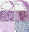
Uveal melanoma (UM) is the most common primary intraocular tumor in adults and usually has a very poor prognosis. Histologically, UMs have been classified in epithelioid cell type, spindle cell type, and mixed cell type. Balloon cells are large pale cells that contain small, hyperchromatic, central nuclei with vesiculated, clear, and lipid-rich cytoplasm. A balloon cell morphology is infrequently observed in naevi and even less frequently in malignant melanomas of the skin, conjunctiva, ciliary body and choroid. In this regard, UMs that exhibit balloon cell features are generally those previously treated with proton beam irradiation and then enucleated, rather than those that directly underwent primary surgery. To the best of our knowledge, very few cases of primary UM showing extensive balloon cell morphology have been reported in scientific literature to date. We herein present an unusual case of primary UM with diffuse balloon cell changes in a 69-year-old woman.
Keywords: balloon cell; eye tumor; non-irradiated melanoma of the choroid; uveal melanoma.
Conflict of interest statement
The authors declare no conflict of interest.
Figures


Similar articles
-
[The prognostic value of uveal melanoma cell type].Arkh Patol. 2021;83(4):14-21. doi: 10.17116/patol20218304114. Arkh Patol. 2021. PMID: 34278756 Russian.
-
Lipoblast-Like Morphology in a Uveal Melanoma with Delayed Metastasis to the Liver.Ocul Oncol Pathol. 2020 Mar;6(2):87-92. doi: 10.1159/000501619. Epub 2019 Sep 4. Ocul Oncol Pathol. 2020. PMID: 32258015 Free PMC article.
-
Intraocular collision tumour: case report and literature review.Graefes Arch Clin Exp Ophthalmol. 2013 May;251(5):1383-8. doi: 10.1007/s00417-012-2216-0. Epub 2012 Dec 12. Graefes Arch Clin Exp Ophthalmol. 2013. PMID: 23232651 Review.
-
Effects of proton beam irradiation on uveal melanomas: a comparative study of Ki-67 expression in irradiated versus non-irradiated melanomas.Br J Ophthalmol. 2000 Jan;84(1):98-102. doi: 10.1136/bjo.84.1.98. Br J Ophthalmol. 2000. PMID: 10611107 Free PMC article.
-
Uveal melanoma: A pathologist's perspective and review of translational developments.Adv Anat Pathol. 2014 Mar;21(2):138-43. doi: 10.1097/PAP.0000000000000010. Adv Anat Pathol. 2014. PMID: 24508696 Review.
Cited by
-
Genetics and RNA Regulation of Uveal Melanoma.Cancers (Basel). 2023 Jan 26;15(3):775. doi: 10.3390/cancers15030775. Cancers (Basel). 2023. PMID: 36765733 Free PMC article. Review.
References
-
- Broggi G., Russo A., Reibaldi M., Russo D., Varricchio S., Bonfiglio V., Spatola C., Barbagallo C., Foti P.V., Avitabile T., et al. Histopathology and Genetic Biomarkers of Choroidal Melanoma. Appl. Sci. 2020;10:8081. doi: 10.3390/app10228081. - DOI
-
- Foti P.V., Travali M., Farina R., Palmucci S., Spatola C., Liardo R.L.E., Milazzotto R., Raffaele L., Salamone V., Caltabiano R., et al. Diagnostic methods and therapeutic options of uveal melanoma with emphasis on MR imaging-Part II: Treatment indications and complications. Insights Imaging. 2021;12:67. doi: 10.1186/s13244-021-01001-w. - DOI - PMC - PubMed
Publication types
LinkOut - more resources
Full Text Sources

