Review
. 2022 Mar 8;12(3):659.
doi: 10.3390/diagnostics12030659.

Anchoring Apparatus of Long Head of the Biceps Tendon: Ultrasonographic Anatomy and Pathologic Conditions

Affiliations
Review

Anchoring Apparatus of Long Head of the Biceps Tendon: Ultrasonographic Anatomy and Pathologic Conditions

Heng Xue et al. Diagnostics (Basel). .

Abstract

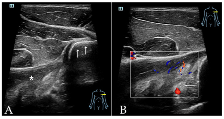
The long head of the biceps tendon (LHBT) has been recognized as an important generator of anterior shoulder pain, causing a significant reduction in the shoulder flexion range. Various tendinous and ligamentous structures form the anchoring apparatus of the LHBT along its course to maintain its appropriate location during shoulder movements, including the coracohumeral ligament (CHL), superior glenohumeral ligament (SGHL), subscapularis (SSC) tendon and supraspinatus (SSP) tendon as well as the less recognized tendons of pectoralis major (PM), latissimus dorsi (LD) and teres major (TM). Lesions of this stabilizing apparatus may lead to an instability of the LHBT, resulting in pain at the anterior shoulder. Ultrasonography (US) has been increasingly used in the assessment of shoulder injuries, including the anchoring apparatus of the LHBT. An accurate diagnosis of these injuries is often challenging, given the complex anatomy and wide spectrum of pathologies. In this review article, US anatomy and common pathologic conditions that affect the anchoring apparatus of the LHBT are discussed, including biceps pulley lesions, adhesive capsulitis, chronic pathology of SSC and SSP tendons, tears in the PM tendon and injuries to the LD and TM. Knowledge of a normal anatomy, an appropriate scanning technique and US findings of common pathologic conditions are the keys to accurate diagnoses.

Keywords: long head of the biceps tendon; rotator cuff; scanning technique; shoulder; ultrasonography.


Conflict of interest statement

The authors declare no conflict of interest.

Figures

Figure 1
Figure 1
(A) Patient position and probe orientation when evaluating rotator interval. (B) US image shows rotator interval. 1 = LHBT, 2 = CHL, 3 = SGHL, 4 = tendon of SSC, 5 = tendon of SSP, 6 = deltoid muscle.
Figure 2
Figure 2
(A) Patient position and probe orientation when evaluating LHBT and bicipital groove. (B) US image shows the LHBT and bicipital groove. White arrows = bicipital groove, 1 = LHBT, 2 = greater tuberosity, 3 = lesser tuberosity, 4 = deltoid muscle.
Figure 3
Figure 3
Schematic drawing of biceps and reflection pulley. 1 = LHBT, 2 = lesser tuberosity, 3 = SSC tendon, 4 = greater tuberosity, 5 = SSP tendon, 6 = SGHL, 7 = CHL, 8 = humeral head, 9 = scapular glenoid, 10 = glenoid labrum. In the drawing, 3, 5 and 7 form the biceps pulley and 6 and the medial band of 7 form the reflection pulley.
Figure 4
Figure 4
(A) Photographs illustrates the patient position and US transducer orientation when evaluating SSC tendon. (B) Photograph illustrates the patient position and US transducer orientation when evaluating SSP tendon. (C) US image shows long axis of SSC tendon. 1 = SSC tendon, 2 = lesser tuberosity, 3 = deltoid muscle, 4 = subdeltoid bursae. (D) US image shows short axis of SSC tendon. (E) US image shows long axis of SSP tendon. (F) US image shows short axis of SSP tendon. 1 = SSP tendon, 2 = greater tuberosity, 3 = humeral head, 4 = deltoid muscle. Arrow = anatomical neck of humerus.
Figure 4
Figure 4
(A) Photographs illustrates the patient position and US transducer orientation when evaluating SSC tendon. (B) Photograph illustrates the patient position and US transducer orientation when evaluating SSP tendon. (C) US image shows long axis of SSC tendon. 1 = SSC tendon, 2 = lesser tuberosity, 3 = deltoid muscle, 4 = subdeltoid bursae. (D) US image shows short axis of SSC tendon. (E) US image shows long axis of SSP tendon. (F) US image shows short axis of SSP tendon. 1 = SSP tendon, 2 = greater tuberosity, 3 = humeral head, 4 = deltoid muscle. Arrow = anatomical neck of humerus.
Figure 5
Figure 5
Transverse US image shows an echogenic linear PM tendon (white arrows), which attaches to the lateral lip aspect of the of the bicipital groove. 1 = biceps muscle, 2 = coracobrachialis muscle, 3 = deltoid muscle.
Figure 6
Figure 6
With the arm of the patient maximally externally rotated (A), transverse US image (B) shows tendons of the LD and TM as echogenic linear structures (white arrows indicate LD tendon and white arrow head indicates TM tendon). Black asterisk = PM tendon, 1 = biceps muscle, 2 = coracobrachialis muscle, 3 = deltoid muscle, 4 = LD muscle. Note that the muscle belly of the LD is significantly hypoechoic due to anisotropic artifact.
Figure 7
Figure 7
The patient was a 50-year-old female, complaining of right shoulder pain and restricted range of movement for 2 months. Transverse US over the bicipital groove demonstrated medially dislocated LHBT, bony irregularities of humerus and swelling of the biceps pulley with hyperemia. A biceps pulley lesion was confirmed with arthroscopic surgery.
Figure 8
Figure 8
US images of a patient with AC. The patient was a 62-year-old female with painful restricted motion of the right shoulder for 6 months. The clinical diagnosis was AC. (A) Transverse image of rotator interval showed increased vascularity. (B) Transverse image obtained slightly distally showed increased fluid in the sheath of LHBT resulting from decreased capsular volume of the shoulder joint. (C) Axillary recess capsule was significantly thickened (between calipers measuring 0.5 cm) compared with the asymptomatic side. (D) Posterior recess capsule was also thickened (between calipers measuring 0.35 cm).
Figure 8
Figure 8
US images of a patient with AC. The patient was a 62-year-old female with painful restricted motion of the right shoulder for 6 months. The clinical diagnosis was AC. (A) Transverse image of rotator interval showed increased vascularity. (B) Transverse image obtained slightly distally showed increased fluid in the sheath of LHBT resulting from decreased capsular volume of the shoulder joint. (C) Axillary recess capsule was significantly thickened (between calipers measuring 0.5 cm) compared with the asymptomatic side. (D) Posterior recess capsule was also thickened (between calipers measuring 0.35 cm).
Figure 9
Figure 9
A 63-year-old male patient, complaining of left shoulder pain and palpable snap when the arm was externally rotated. (A) Long axis of the SSP tendon showed full thickness tears in the anterior border (white arrow). (B) With the arm externally rotated, the short axis of the LHBT showed dislocation of the tendon to the medial side of the lesser tuberosity, superficial to the SSC tendon. (1 = LHBT, 2 = lesser tuberosity, 3 = SSC tendon).
Figure 10
Figure 10
PM tendon tears in 2 patients. (A) 64-year-old male patient, complaining of focal pain and weakness with arm adduction after swinging from a high bar. US showed the absence of the PM tendon and retraction of PM muscle fibers. (B) 25-year-old male patient, complaining of immediate pain after a bench press exercise. US showed retracted PM muscle stump and surrounding anechoic fluid.
Figure 11
Figure 11
LD muscle belly strains in a 34-year-old professional bodybuilder. He presented with acute and focal pain in the back of the left shoulder after injury. (A) Transverse image of the LD tendon (white arrows) showed swelling and disrupted fibrillar pattern of the muscle belly (white asterisk). (B) The swelling in the LD muscle belly showed increased vascularity. As the tendon of LD was intact, conservative treatment was recommended.

Similar articles

Cited by

References

    1. Elser F., Braun S., Dewing C.B., Giphart J.E., Millett P.J. Anatomy, function, injuries, and treatment of the long head of the biceps brachii tendon. Arthroscopy. 2011;27:581–592. doi: 10.1016/j.arthro.2010.10.014. - DOI - PubMed
    1. Zappia M., Reginelli A., Russo A., D’Agosto G.F., Di Pietto F., Genovese E.A., Coppolino F., Brunese L. Long head of the biceps tendon and rotator interval. Musculoskelet Surg. 2013;97((Suppl. 2)):S99–S108. doi: 10.1007/s12306-013-0290-z. - DOI - PubMed
    1. Mellano C.R., Shin J.J., Yanke A.B., Verma N.N. Disorders of the long head of the biceps tendon. Instr Course Lect. 2015;64:567–576. - PubMed
    1. Morag Y., Bedi A., Jamadar D.A. The rotator interval and long head biceps tendon: Anatomy, function, pathology, and magnetic resonance imaging. Magn. Reson. Imaging Clin. N. Am. 2012;20:229–259. doi: 10.1016/j.mric.2012.01.012. - DOI - PubMed
    1. Werner A., Mueller T., Boehm D., Gohlke F. The stabilizing sling for the long head of the biceps tendon in the rotator cuff interval. A histoanatomic study. Am. J. Sports Med. 2000;28:28–31. doi: 10.1177/03635465000280011701. - DOI - PubMed