

The Importance of Cardiac Computed Tomography in the Diagnosis of Caseous Calcification of the Mitral Annulus-Case Reports
- PMID: 35328220
- PMCID: PMC8947161
- DOI: 10.3390/diagnostics12030667
The Importance of Cardiac Computed Tomography in the Diagnosis of Caseous Calcification of the Mitral Annulus-Case Reports
Abstract
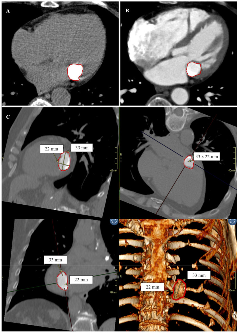
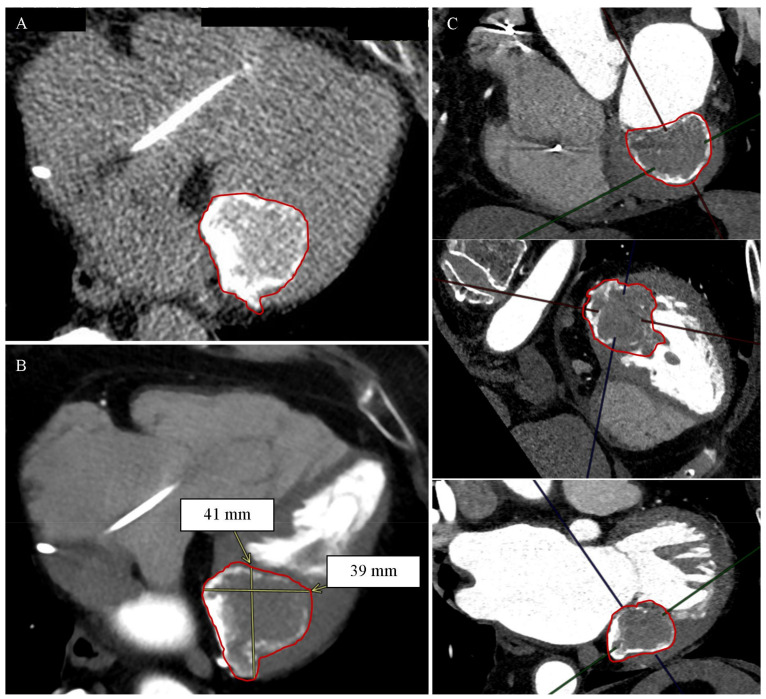
Mitral annular calcification (MAC) is a common pathology of the mitral valve. In rare cases, calcifications occur in the mitral annulus degenerate serous; the caseous calcification of the mitral annulus (CCMA) then develops. Detection of CCMA is often random and requires differentiation from heart tumors or an abscess. The paper presents two cases of patients with ambiguous focal lesions of the mitral valve in echocardiography. In the first case, the cardiac computed tomography (CCT) showed a spherical, slightly irregular structure measuring approximately 33 × 22 mm, which was in contact with the posterior mitral valve leaflet from the lumen of the left ventricle. The lesion was heterogeneously intense, with an average density of about 500 HU and up to 975 HU on the periphery; it was not enhanced after the administration of a contrast agent. In the second case, the CCT revealed a heterogeneous, highly calcified structure in the peripheral zone and intermediate density in the central zone in the topography of the posterior mitral valve leaf, with dimensions up to about 41 × 31 mm in the plane of the valve leaflet, passing into the lumen of the left ventricle along its inferolateral wall to a depth of about 3.5 cm. In both cases, CCT enabled the diagnosis of CCMA. In conclusion, cardiac computed tomography may be decisive in the case of suspected caseous calcification of the mitral annulus where there is ambiguous echocardiography.
Keywords: cardiac computed tomography; caseous calcification of the mitral annulus; mitral annular calcification.
Conflict of interest statement
The authors declare no conflict of interest.
Figures
Similar articles
-
Surgery for mitral annular caseous calcification-related calcified amorphous tumor: a case report.Gen Thorac Cardiovasc Surg Cases. 2023 May 15;2(1):21. doi: 10.1186/s44215-023-00042-5. Gen Thorac Cardiovasc Surg Cases. 2023. PMID: 39516942 Free PMC article.
-
An extensive caseous calcification of the mitral annulus complicated with severe mitral regurgitation.Echocardiography. 2018 Feb;35(2):282-284. doi: 10.1111/echo.13800. Epub 2018 Jan 18. Echocardiography. 2018. PMID: 29346710
-
Ruptured caseous calcification of the mitral annulus.Australas J Ultrasound Med. 2020 Dec 20;24(2):106-111. doi: 10.1002/ajum.12238. eCollection 2021 May. Australas J Ultrasound Med. 2020. PMID: 34765419 Free PMC article.
-
Caseous calcification of the mitral annulus: a review.Clin Cardiol. 2013 Oct;36(10):E27-31. doi: 10.1002/clc.22199. Epub 2013 Aug 27. Clin Cardiol. 2013. PMID: 24038099 Free PMC article. Review.
-
Mitral Annular Calcification-Related Valvular Disease: A Challenging Entity.J Clin Med. 2024 Feb 3;13(3):896. doi: 10.3390/jcm13030896. J Clin Med. 2024. PMID: 38337590 Free PMC article. Review.
References
Publication types
LinkOut - more resources
Full Text Sources
