Use of a FluoroType® System for the Rapid Detection of Patients with Multidrug-Resistant Tuberculosis-State of the Art Case Presentations
- PMID: 35328264
- PMCID: PMC8947722
- DOI: 10.3390/diagnostics12030711
Use of a FluoroType® System for the Rapid Detection of Patients with Multidrug-Resistant Tuberculosis-State of the Art Case Presentations
Abstract
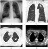
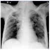
According to the World Health Organization (WHO), there were 465,000 cases of tuberculosis caused by strains resistant to at least two first-line anti-tuberculosis drugs: rifampicin and isoniazid (MDR-TB). In light of the growing problem of drug resistance in Mycobacterium tuberculosis across laboratories worldwide, the rapid identification of drug-resistant strains of the Mycobacterium tuberculosis complex poses the greatest challenge. Progress in molecular biology and the development of nucleic acid amplification assays have paved the way for improvements to methods for the direct detection of Mycobacterium tuberculosis in specimens from patients. This paper presents two cases that illustrate the implementation of molecular tools in the recognition of drug-resistant tuberculosis.
Keywords: FluoroType MTBDR; Mycobacterium tuberculosis; drug resistance; isoniazid; molecular; rifampin; tuberculosis.
Conflict of interest statement
The authors declare no conflict of interest.
Figures


Similar articles
-
Diagnostic Accuracy and Utility of FluoroType MTBDR, a New Molecular Assay for Multidrug-Resistant Tuberculosis.J Clin Microbiol. 2018 Aug 27;56(9):e00531-18. doi: 10.1128/JCM.00531-18. Print 2018 Sep. J Clin Microbiol. 2018. PMID: 29976588 Free PMC article.
-
Validation of the FluoroType MTBDR Assay for Detection of Rifampin and Isoniazid Resistance in Mycobacterium tuberculosis Complex Isolates.J Clin Microbiol. 2018 May 25;56(6):e00072-18. doi: 10.1128/JCM.00072-18. Print 2018 Jun. J Clin Microbiol. 2018. PMID: 29593055 Free PMC article.
-
API TB Consensus Guidelines 2006: Management of pulmonary tuberculosis, extra-pulmonary tuberculosis and tuberculosis in special situations.J Assoc Physicians India. 2006 Mar;54:219-34. J Assoc Physicians India. 2006. PMID: 16800350
-
Direct susceptibility testing for multi drug resistant tuberculosis: a meta-analysis.BMC Infect Dis. 2009 May 20;9:67. doi: 10.1186/1471-2334-9-67. BMC Infect Dis. 2009. PMID: 19457256 Free PMC article. Review.
-
Management of Multidrug-Resistant Tuberculosis.Semin Respir Crit Care Med. 2018 Jun;39(3):310-324. doi: 10.1055/s-0038-1661383. Epub 2018 Aug 2. Semin Respir Crit Care Med. 2018. PMID: 30071546 Review.
Cited by
-
New insight in molecular detection of Mycobacterium tuberculosis.AMB Express. 2024 Jun 21;14(1):74. doi: 10.1186/s13568-024-01730-3. AMB Express. 2024. PMID: 38907086 Free PMC article. Review.
References
-
- Global Tuberculosis Report 2020. World Health Organization; Geneva, Switzerland: 2020. Licence: CC BY-NC-SA 3.0 IGO.
-
- European Centre for Disease Prevention and Control . Handbook on Tuberculosis Laboratory Diagnostic Methods in the European Union–Updated 2018. ECDC; Stockholm, Sweden: 2018.
-
- Korzeniewska-Koseła M., editor. Tuberculosis and Respiratory Tract Diseases in Poland in 2019. Institute of Tuberculosis and Lung Diseases; Warsaw, Poland: 2020.
-
- Antonenka U., Hofmann-Thiel S., Turaev L., Esenalieva A., Abdulloeva M., Sahalchyk E., Alnour T., Hoffmann H. Coparison of Xpert MTB/RIF with ProbeTec ET DTB and COBAS TaqMan MTB for direct detection of M. tuberculosis complex in respiratory specimens. BMC Infect. Dis. 2013;13:280. doi: 10.1186/1471-2334-13-280. - DOI - PMC - PubMed
Publication types
LinkOut - more resources
Full Text Sources

