. 2022 Mar 15;12(3):712.
doi: 10.3390/diagnostics12030712.

Commercially Available Heart Rate Monitor Repurposed for Automatic Arrhythmia Detection with Snapshot Electrocardiographic Capability: A Pilot Validation

Affiliations

Commercially Available Heart Rate Monitor Repurposed for Automatic Arrhythmia Detection with Snapshot Electrocardiographic Capability: A Pilot Validation

Chiara Martini et al. Diagnostics (Basel). .

Abstract

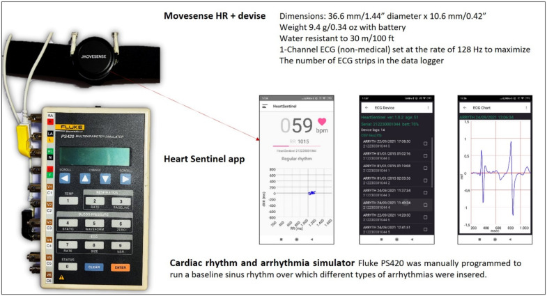
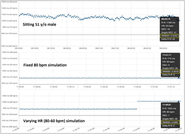
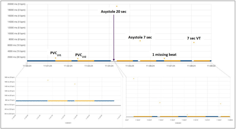
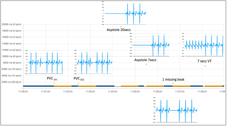
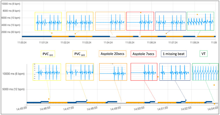
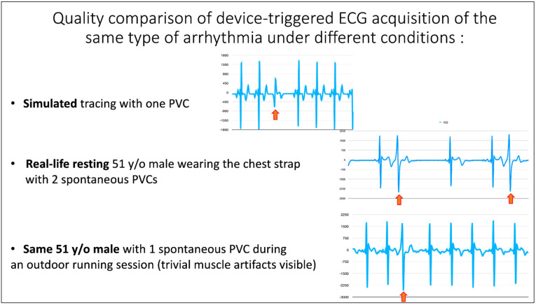
The usefulness of opportunistic arrhythmia screening strategies, using an electrocardiogram (ECG) or other methods for random "snapshot" assessments is limited by the unexpected and occasional nature of arrhythmias, leading to a high rate of missed diagnosis. We have previously validated a cardiac monitoring system for AF detection pairing simple consumer-grade Bluetooth low-energy (BLE) heart rate (HR) sensors with a smartphone application (RITMIA™, Heart Sentinel srl, Italy). In the current study, we test a significant upgrade to the above-mentioned system, thanks to the technical capability of new HR sensors to run algorithms on the sensor itself and to acquire, and store on-board, single-lead ECG strips. We have reprogrammed an HR monitor intended for sports use (Movensense HR+) to run our proprietary RITMIA algorithm code in real-time, based on RR analysis, so that if any type of arrhythmia is detected, it triggers a brief retrospective recording of a single-lead ECG, providing tracings of the specific arrhythmia for later consultation. We report the initial data on the behavior, feasibility, and high diagnostic accuracy of this ultra-low weight customized device for standalone automatic arrhythmia detection and ECG recording, when several types of arrhythmias were simulated under different baseline conditions. Conclusions: The customized device was capable of detecting all types of simulated arrhythmias and correctly triggered a visually interpretable ECG tracing. Future human studies are needed to address real-life accuracy of this device.

Keywords: arrhythmia; cardiac monitoring; cardiovascular prevention; digital cardiology; electrocardiogram; heart failure; sensors; sports cardiology.


Conflict of interest statement

The authors declare no conflict of interest.

Figures

Figure 1
Figure 1
The simulation system components, the Movesense HR+ sensor attached to the chest-belt, and in parallel, wired to the output of a Fluke PS420 simulator. In the middle, three screenshots from the Heart Sentinel app are shown, from left to right, demonstrating: A real-time RR plot, a log of the ECG strips recorded, and a sample of 5-s ECG strip triggered by a premature ventricular beat, obtained after clicking on one of the events in the log. The Heart Sentinel app (in this case running on an Android smartphone) was used during simulations for continuous upload of RR intervals to the cloud, to be later shown in the back-office cloud app matched with stored ECG tracings.
Figure 2
Figure 2
While the physiological heart rate variability extrinsicates in an RMSSD index around 0.025 in an adult male in the sitting position in «real-life» (top), the simulation with a fixed heart rate (middle) does not account for physiological RR variability (RMSSD 0.0078), while varying heart rate between 80 bpm and 60 bpm in the simulation brings the RMSSD index back to the order of magnitude of physiological values (RMSSD 0.0367) (bottom).
Figure 3
Figure 3
PVC, premature ventricular complex; VT, ventricular tachycardia. Beats considered from normal sinus rhythm are automatically labeled in blue, while non-AF arrhythmia in orange. If AF is present, it is labeled in red. After a single PVC or any other abnormal beat (or pause) recognized as an arrhythmic event by the algorithm, a given number of subsequent beats remain labeled in orange until the last arrhythmic beat exits the moving matrix made by a fixed number of consecutive prior beats, used for computation of variability in real-time. Labeled beats always follow the arrhythmic event, but they can trigger the acquisition of other electrocardiograms only once the rhythm reverts to normal (at least one blue beat).
Figure 4
Figure 4
PVC, premature ventricular complex. Beats considered from normal sinus rhythm are automatically labeled in blue, while non-AF arrhythmia in orange. After a single PVC or any other abnormal beat (or pause) recognized as an arrhythmic event by the algorithm, a given number of subsequent beats remain labeled in orange until the last arrhythmic beat exits the moving matrix made by a fixed number of consecutive prior beats, used for computation of variability in real-time. Labeled beats always follow the arrhythmic event, but they can trigger the acquisition of other electrocardiograms only once the rhythm re-verts to normal (at least one blue beat). The missing beat is also recognized.
Figure 5
Figure 5
PVC, premature ventricular complex. Beats considered from normal sinus rhythm are automatically labeled in blue, while non-AF arrhythmia in orange. After a single PVC or any other abnormal beat (or pause) recognized as an arrhythmic event by the algorithm, a given number of subsequent beats remain labeled in orange until the last arrhythmic beat exits the moving matrix made by a fixed number of consecutive prior beats, used for computation of variability in real-time. Labeled beats always follow the arrhythmic event, but they can trigger the acquisition of other electrocardiograms only once the rhythm re-verts to normal (at least one blue beat). The missing beat is also recognized.
Figure 6
Figure 6
ECG, electrocardiogram; PVC, premature ventricular complex. Quality comparison of device-triggered ECG acquisition of the same type of arrhythmia under different conditions of rest and stress.

References

    1. Noseworthy P.A., Kaufman E.S., Chen L.Y., Chung M.K., Elkind M.S., Joglar J.A., Leal M.A., McCabe P.J., Pokorney S.D., Yao X. Subclinical and Device-Detected Atrial Fibrillation: Pondering the Knowledge Gap: A Scientific Statement From the American Heart Association. Circulation. 2019;140:e944–e963. doi: 10.1161/CIR.0000000000000740. - DOI - PMC - PubMed
    1. Svendsen J.H., Diederichsen S.Z., Højberg S., Krieger D.W., Graff C., Kronborg C., Olesen M.S., Nielsen J.B., Holst A.G., Brandes A., et al. Implantable loop recorder detection of atrial fibrillation to prevent stroke (The LOOP Study): A randomised controlled trial. Lancet. 2021;398:1507–1516. doi: 10.1016/S0140-6736(21)01698-6. - DOI - PubMed
    1. Sana F., Isselbacher E.M., Singh J.P., Heist E.K., Pathik B., Armoundas A.A. Wearable Devices for Ambulatory Cardiac Monitoring. J. Am. Coll. Cardiol. 2020;75:1582–1592. doi: 10.1016/j.jacc.2020.01.046. - DOI - PMC - PubMed
    1. McConnell M.V., Turakhia M.P., Harrington R.A., King A.C., Ashley E.A. Mobile Health Advances in Physical Activity, Fitness, and Atrial Fibrillation. J. Am. Coll. Cardiol. 2018;71:2691–2701. doi: 10.1016/j.jacc.2018.04.030. - DOI - PubMed
    1. Zhu H., Pan Y., Wu F., Huan R. Optimized Electrode Locations for Wearable Single-Lead ECG Monitoring Devices: A Case Study Using WFEES Modules based on the LANS Method. Sensors. 2019;19:4458. doi: 10.3390/s19204458. - DOI - PMC - PubMed

LinkOut - more resources