

Use of Gallbladder Width Measurement by Computed Tomography in the Diagnosis of Acute Cholecystitis
- PMID: 35328274
- PMCID: PMC8946906
- DOI: 10.3390/diagnostics12030721
Use of Gallbladder Width Measurement by Computed Tomography in the Diagnosis of Acute Cholecystitis
Abstract
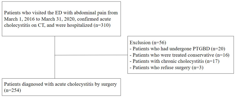
This study aimed to evaluate the diagnostic value of gallbladder width measurement with computed tomography (CT) in patients with acute cholecystitis. This retrospective case−control study was conducted between March 2016 and March 2020 at a tertiary emergency department. Of 310 patients, 254 patients with acute cholecystitis confirmed by surgery were compared with 254 patients diagnosed with other diseases (controls). In the acute cholecystitis group, the number of older patients with underlying illnesses was much higher (64% of men). Upon CT, the median (interquartile range [IQR]) gallbladder width was significantly longer in patients with acute cholecystitis (2.26 [1.82−2.78] cm vs. 3.73 [3.32−4.16] cm, p < 0.001). The optimal cut-off value of gallbladder width for differentiating acute cholecystitis was 3.12 cm, showing a sensitivity of 88% and specificity of 86%. In a multivariable analysis using a logistic regression model for diagnosing acute cholecystitis with CT findings (gallbladder width, length, stone, wall thickening, and pericholecystic fluid), a gallbladder width of ≥3.12 cm was significantly meaningful, even when adjusting for other variables (odds ratio 37.9; p < 0.001). Therefore, an increase in gallbladder width (≥3.12 cm) measured with CT can be a simple and sensitive diagnostic sign of acute cholecystitis, supporting the underlying pathophysiology of bile outflow obstruction.
Keywords: CT; acute cholecystitis; computed tomography; diagnosis; gallbladder.
Conflict of interest statement
The authors declare no conflict of interest.
Figures


Similar articles
-
Pope's hat sign: another valuable CT finding of early acute cholecystitis.Abdom Radiol (NY). 2018 Jul;43(7):1693-1702. doi: 10.1007/s00261-017-1421-z. Abdom Radiol (NY). 2018. PMID: 29198010
-
Differentiation of acute cholecystitis from chronic cholecystitis: Determination of useful multidetector computed tomography findings.Medicine (Baltimore). 2018 Aug;97(33):e11851. doi: 10.1097/MD.0000000000011851. Medicine (Baltimore). 2018. PMID: 30113479 Free PMC article.
-
[Personal experience in 71 consecutive patients with acute cholecystitis].Radiol Med. 2000 Jan-Feb;99(1-2):62-7. Radiol Med. 2000. PMID: 10803189 Italian.
-
Computed tomographic evaluation of gallbladder disease.Crit Rev Diagn Imaging. 1987;27(2):113-52. Crit Rev Diagn Imaging. 1987. PMID: 3301215 Review.
-
Acute cholecystitis: CT findings.Semin Ultrasound CT MR. 2000 Feb;21(1):56-63. doi: 10.1016/s0887-2171(00)90013-1. Semin Ultrasound CT MR. 2000. PMID: 10688067 Review.
Cited by
-
Computed tomography versus ultrasound for the diagnosis of acute cholecystitis: a systematic review and meta-analysis.Eur Radiol. 2024 Nov;34(11):6967-6979. doi: 10.1007/s00330-024-10783-8. Epub 2024 May 17. Eur Radiol. 2024. PMID: 38758253
-
Ultrasound of the Gallbladder-An Update on Measurements, Reference Values, Variants and Frequent Pathologies: A Scoping Review.Life (Basel). 2025 Jun 11;15(6):941. doi: 10.3390/life15060941. Life (Basel). 2025. PMID: 40566593 Free PMC article. Review.
References
-
- Kimura Y., Takada T., Kawarada Y., Nimura Y., Hirata K., Sekimoto M., Yoshida M., Mayumi T., Wada K., Miura F., et al. Definitions, pathophysiology, and epidemiology of acute cholangitis and cholecystitis: Tokyo Guidelines. J. Hepatobiliary Pancreat. Surg. 2007;14:15–26. doi: 10.1007/s00534-006-1152-y. - DOI - PMC - PubMed
-
- de Burlet K., Lam A., Larsen P., Dennett E. Acute abdominal pain-changes in the way we assess it over a decade. N. Z. Med. J. 2017;130:39–44. - PubMed
-
- Yokoe M., Hata J., Takada T., Strasberg S.M., Asbun H.J., Wakabayashi G., Kozaka K., Endo I., Deziel D.J., Miura F., et al. Tokyo Guidelines 2018: Diagnostic criteria and severity grading of acute cholecystitis (with videos) J. Hepatobiliary Pancreat. Sci. 2018;25:41–54. doi: 10.1002/jhbp.515. - DOI - PubMed
LinkOut - more resources
Full Text Sources
