Review
. 2022 Mar 19;12(3):753.
doi: 10.3390/diagnostics12030753.

The Utility of Endoscopic-Ultrasonography-Guided Tissue Acquisition for Solid Pancreatic Lesions

Affiliations
Review

The Utility of Endoscopic-Ultrasonography-Guided Tissue Acquisition for Solid Pancreatic Lesions

Hiroki Tanaka et al. Diagnostics (Basel). .

Abstract

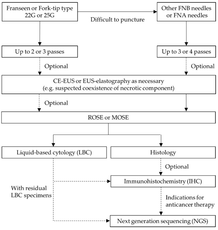
Endoscopic-ultrasonography-guided tissue acquisition (EUS-TA) has been widely performed for the definitive diagnosis of solid pancreatic lesions (SPLs). As the puncture needles, puncture techniques, and sample processing methods have improved, EUS-TA has shown higher diagnostic yields and safety. Recently, several therapeutic target genomic biomarkers have been clarified in pancreatic ductal carcinoma (PDAC). Although only a small proportion of patients with PDAC can benefit from precision medicine based on gene mutations at present, precision medicine will also be further developed for SPLs as more therapeutic target genomic biomarkers are identified. Advances in next-generation sequencing (NGS) techniques enable the examination of multiple genetic mutations in limited tissue samples. EUS-TA is also useful for NGS and will play a more important role in determining treatment strategies. In this review, we describe the utility of EUS-TA for SPLs.

Keywords: EUS-guided fine-needle aspiration; EUS-guided fine-needle biopsy; EUS-guided tissue acquisition; endoscopic ultrasonography; pancreatic ductal adenocarcinoma; pancreatic neuroendocrine neoplasms; solid pancreatic lesions.


Conflict of interest statement

The authors declare no conflict of interest.

Figures

Figure 1
Figure 1
Anaplastic carcinoma of the pancreas. (a) Contrast-enhanced computed tomography (CE-CT) demonstrated a poorly circumscribed, low-density mass lesion at the pancreatic head concomitant with chronic pancreatitis (arrow). (b) Multiplanar reconstruction (MPR) of CE-CT image. (c) EUS-FNA was performed using a Menghini-type 22-gause needle with the fanning technique, and negative pressure was applied with a 20 mL syringe. (d) Histology of the specimens by EUS-FNA showed osteoclastic polynuclear giant cells. (hematoxylin and eosin staining, ×400).
Figure 2
Figure 2
Metastatic renal cell carcinoma to the pancreas. (a) Endoscopic ultrasonography (EUS) revealed a well-circumscribed, homogenous low-echoic mass lesion 17 mm in size at the pancreatic tail (arrow). (b) EUS-FNA was performed using a Franseen-type 22-gause needle with the fanning technique, and negative pressure was applied with a 20 mL syringe. (cf) Hematoxylin and eosin staining showed tumor cells with clear cytoplasm (c). In immunohistochemistry, the tumor cells were positive for CD10 (d), PAX8 (e) and Vimentin (f).
Figure 2
Figure 2
Metastatic renal cell carcinoma to the pancreas. (a) Endoscopic ultrasonography (EUS) revealed a well-circumscribed, homogenous low-echoic mass lesion 17 mm in size at the pancreatic tail (arrow). (b) EUS-FNA was performed using a Franseen-type 22-gause needle with the fanning technique, and negative pressure was applied with a 20 mL syringe. (cf) Hematoxylin and eosin staining showed tumor cells with clear cytoplasm (c). In immunohistochemistry, the tumor cells were positive for CD10 (d), PAX8 (e) and Vimentin (f).
Figure 3
Figure 3
Metastatic bladder carcinoma to the pancreas. (a) Endoscopic ultrasonography (EUS) revealed a well-circumscribed, homogenous low-echoic mass lesion 8 mm in size at the pancreatic tail (arrow). (b) EUS-FNA was performed using a Franseen-type 22-gause needle, with negative pressure applied with a 20 mL syringe. The specimen included white core tissue, red core tissue, and a liquid component. (c) Hematoxylin and eosin staining findings were suspicious for urothelial cell carcinoma. (d) The tumor cells were positive for GATA3.
Figure 4
Figure 4
Our recommendations for EUS-FNA of solid pancreatic lesions based on this review.

Similar articles

Cited by

References

    1. Hewitt M.J., McPhail M.J., Possamai L., Dhar A., Vlavianos P., Monahan K.J. EUS-guided FNA for diagnosis of solid pancreatic neoplasms: A meta-analysis. Gastrointest. Endosc. 2012;75:319–331. doi: 10.1016/j.gie.2011.08.049. - DOI - PubMed
    1. Facciorusso A., Bajwa H.S., Menon K., Buccino V.R., Muscatiello N. Comparison between 22G aspiration and 22G biopsy needles for EUS-guided sampling of pancreatic lesions: A meta-analysis. Endosc. Ultrasound. 2020;9:167–174. doi: 10.4103/eus.eus_4_19. - DOI - PMC - PubMed
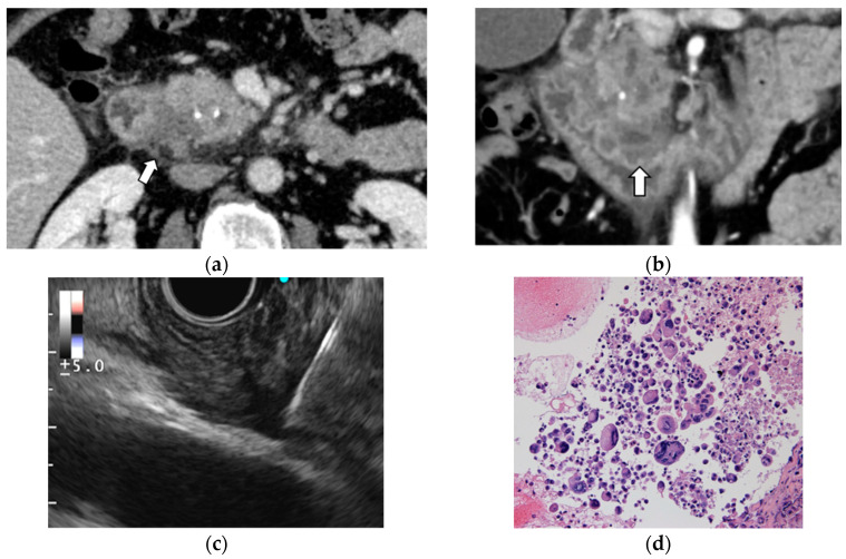
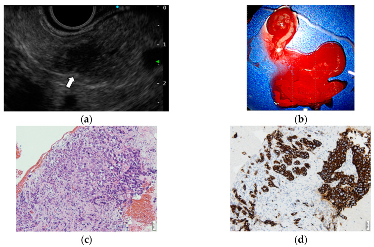
    1. Yoshida N., Kanno A., Masamune A., Nabeshima T., Hongo S., Miura S., Takikawa T., Hamada S., Kikuta K., Kume K., et al. Pancreatic Acinar Cell Carcinoma with Multiple Liver Metastases Effectively Treated by S-1 Chemotherapy. Intern. Med. 2018;57:3529–3535. doi: 10.2169/internalmedicine.0294-17. - DOI - PMC - PubMed
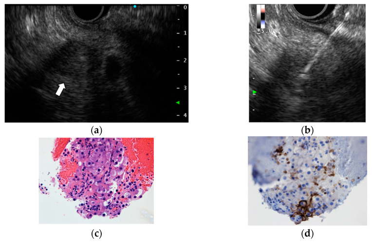
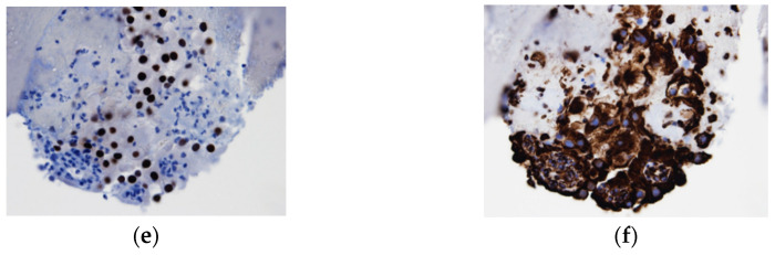
    1. Oka K., Inoue K., Sugino S., Harada T., Tsuji T., Nakashima S., Katayama T., Okuda T., Kin S., Nagata A., et al. Anaplastic carcinoma of the pancreas diagnosed by endoscopic ultrasound-guided fine-needle aspiration: A case report and review of the literature. J. Med. Case Rep. 2018;12:152. doi: 10.1186/s13256-018-1615-1. - DOI - PMC - PubMed
    1. Khashab M.A., Emerson R.E., DeWitt J.M. Endoscopic ultrasound-guided fine-needle aspiration for the diagnosis of anaplastic pancreatic carcinoma: A single-center experience. Pancreas. 2010;39:88–91. doi: 10.1097/MPA.0b013e3181bba268. - DOI - PubMed