

Two-Photon Vision in Age-Related Macular Degeneration: A Translational Study
- PMID: 35328313
- PMCID: PMC8947013
- DOI: 10.3390/diagnostics12030760
Two-Photon Vision in Age-Related Macular Degeneration: A Translational Study
Abstract
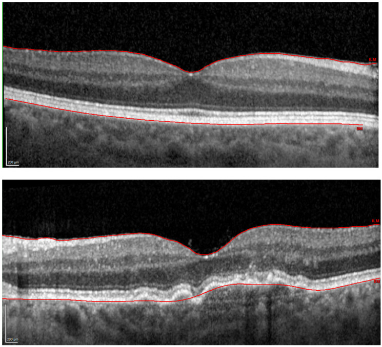
The recently introduced term “two-photon vision” relates to the visual perception resulting from a simultaneous absorption of two photons by photoreceptors. In this study, we determined two-photon retinal sensitivity in age-related macular degeneration (AMD) and compared it that in normal aging. Microperimetry was performed with visible (white) light and infrared (IR) light, which was perceived as green in the two-photon stimulation. In total, 45 subjects were included with one (better) eye studied. Furthermore, best-corrected visual acuity (VA) and ocular straylight were assessed. AMD resulted in decreased median (interquartile range) logMAR VA, i.e., 0.15 (0.05; 0.24), which in normal eyes was −0.02 (−0.06; 0.02). The two groups showed comparable straylight levels. Sensitivity to IR light was significantly lower in the AMD group (p < 0.001): 8.3 (7.4, 9.3) dB than in controls 10.7 (9.7, 11.2) dB. AMD also significantly affected visible light sensitivity (p < 0.001): 14.0 (11.0; 15.5) dB vs. 18.0 (16.3; 18.9) dB. Notably, the two-photon approach yielded a lower data spread. In conclusion, AMD considerably impairs retinal sensitivity measured in the single- and two-photon realm. However, two-photon-vision microperimetry may improve the testing accuracy and offer an additional diagnostic parameter (beyond VA measurements) for retinal function assessment.
Keywords: AMD; microperimetry; normal aging; retinal sensitivity; two-photon vision.
Conflict of interest statement
K.K. is a co-author of U.S. patent 10856734 owned by Polgenix Inc. for “Systems and methods of infrared psychophysical measurement”. G.Ł., A.Z., L.K., A.R., R.K. and G.U.A. declare no conflict of interest.
Figures


Similar articles
-
Correlation of macular sensitivity measures and visual acuity to vision-related quality of life in patients with age-related macular degeneration.BMC Ophthalmol. 2021 Mar 23;21(1):149. doi: 10.1186/s12886-021-01901-x. BMC Ophthalmol. 2021. PMID: 33757447 Free PMC article.
-
Longitudinal changes in microperimetry and low luminance visual acuity in age-related macular degeneration.JAMA Ophthalmol. 2015 Apr;133(4):442-8. doi: 10.1001/jamaophthalmol.2014.5963. JAMA Ophthalmol. 2015. PMID: 25632841
-
Clinical Application of Infrared-Light Microperimetry in the Assessment of Scotopic-Eye Sensitivity.Transl Vis Sci Technol. 2020 Jul 7;9(8):7. doi: 10.1167/tvst.9.8.7. eCollection 2020 Jul. Transl Vis Sci Technol. 2020. PMID: 32855854 Free PMC article.
-
Photobiomodulation for non-exudative age-related macular degeneration.Cochrane Database Syst Rev. 2021 May 6;5(5):CD013029. doi: 10.1002/14651858.CD013029.pub2. Cochrane Database Syst Rev. 2021. PMID: 34097768 Free PMC article.
-
Radiotherapy for neovascular age-related macular degeneration.Cochrane Database Syst Rev. 2020 Aug 26;8(8):CD004004. doi: 10.1002/14651858.CD004004.pub4. Cochrane Database Syst Rev. 2020. PMID: 32844399 Free PMC article.
Cited by
-
From mouse to human: Accessing the biochemistry of vision in vivo by two-photon excitation.Prog Retin Eye Res. 2023 Mar;93:101170. doi: 10.1016/j.preteyeres.2023.101170. Epub 2023 Feb 12. Prog Retin Eye Res. 2023. PMID: 36787681 Free PMC article. Review.
-
Laser pulse train parameters determine the brightness of a two-photon stimulus.Biomed Opt Express. 2023 May 24;14(6):2857-2872. doi: 10.1364/BOE.489890. eCollection 2023 Jun 1. Biomed Opt Express. 2023. PMID: 37342710 Free PMC article.
-
Method for the determination of the luminance of two-photon vision stimuli.Biomed Opt Express. 2024 Sep 9;15(10):5818-5830. doi: 10.1364/BOE.525180. eCollection 2024 Oct 1. Biomed Opt Express. 2024. PMID: 39421759 Free PMC article.
References
-
- CIE . Commission Internationale de L’eclairage Proceedings, 1931. Cambridge University; Cambridge, UK: 1932.
-
- Palczewska G., Vinberg F., Stremplewski P., Bircher M.P., Salom D., Komar K., Zhang J., Cascella M., Wojtkowski M., Kefalov V.J., et al. Human infrared vision is triggered by two-photon chromophore isomerization. Proc. Natl. Acad. Sci. USA. 2014;111:E5445–E5454. doi: 10.1073/pnas.1410162111. - DOI - PMC - PubMed
Grants and funding
LinkOut - more resources
Full Text Sources
Miscellaneous
