Review
. 2022 Mar 21;12(3):763.
doi: 10.3390/diagnostics12030763.

Overview of Lung Ultrasound in Pediatric Cardiology

Affiliations
Review

Overview of Lung Ultrasound in Pediatric Cardiology

Massimiliano Cantinotti et al. Diagnostics (Basel). .

Abstract

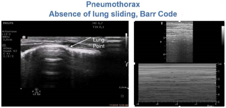
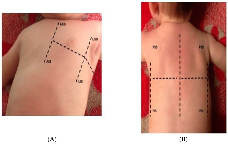
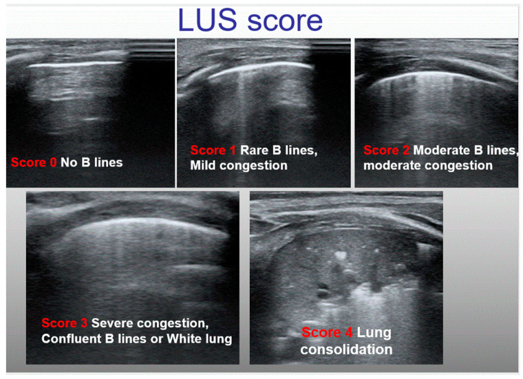
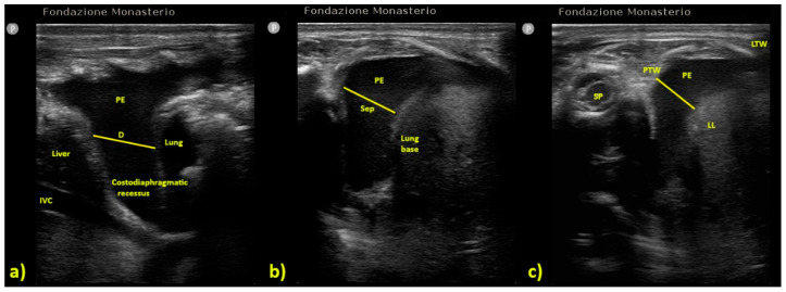
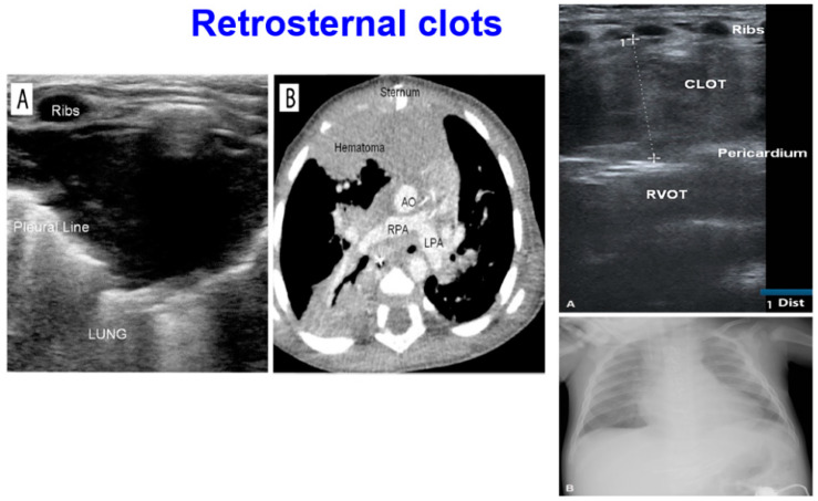
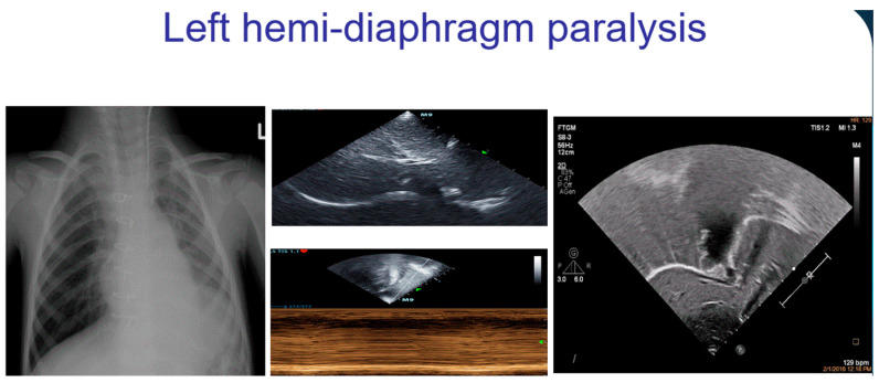
Lung ultrasound (LUS) is increasing in its popularity for the diagnosis of pulmonary complications in acute pediatric care settings. Despite the high incidence of pulmonary complications for patients with pediatric cardiovascular and congenital heart disease, especially in children undergoing cardiac surgery, the use of LUS remains quite limited in these patients. The aim of this review is to provide a comprehensive overview and list of current potential applications for LUS in children with congenital heart disease, post-surgery. We herein describe protocols for LUS examinations in children, discuss diagnostic criteria, and introduce methods for the diagnosis and classification of pulmonary disease commonly encountered in pediatric cardiology (e.g., pleural effusion, atelectasis, interstitial edema, pneumothorax, pneumonia, and diaphragmatic motion analysis). Furthermore, applications of chest ultrasounds for the evaluation of the retrosternal area, and in particular, systematic search criteria for retrosternal clots, are illustrated. We also discussed the potential applications of LUS, including the guidance of interventional procedures, namely lung recruitment and drainage insertion. Lastly, we analyzed current gaps in knowledge, including the difficulty of the quantification of pleural effusion and atelectasis, and the need to differentiate different etiologies of B-lines. We concluded with future applications of LUS, including strain analysis and advanced analysis of diaphragmatic mechanics. In summary, US is an easy, accurate, fast, cheap, and radiation-free tool for the diagnosis and follow-up of major pulmonary complications in pediatric cardiac surgery, and we strongly encourage its use in routine practice.

Keywords: cardiac; congenital; echo; pediatric; ultrasound.


Conflict of interest statement

The authors declare no conflict of interest.

Figures

Figure 4
Figure 4
And Video S1: Diagnosis of pneumothorax. The lung pointy (e.g., the point where the pleura stops its movement) is highlighted on the left side. On the right side, the typical Barcode sign on M-mode is shown.
Figure 1
Figure 1
Six segments score. Each hemithorax is divided into 3 major quadrants (anterior, lateral, and posterior). Each quadrant is further subdivided into the upper and the lower half. (A) anterior and lateral, (B) posterior quadrants.
Figure 2
Figure 2
An example of semiquantitative LUS score that we have recently validated.
Figure 3
Figure 3
Formulas for pleural effusion quantification according to different authors: (a) Usta et al. [24], PEV is calculated by the formula D (mm) × 16, (b) Balik et al. [23] PEV is calculated by the formula Sep (mm) × 20; (c) Eibenberger [25] the major effusion’s diameter (D) is associated with PEV on a progressive scale (e.g., 10 mm correspond to 50–300 mL of PEV, 20 mm to 150–310 mL, etc.). (Table 2) D = distance; IVC = Inferior Vena Cava; LL = Lung Lower Lobe; LTW = Lateral Thoracic Wall; PE = Pleural Effusion; PEV = Pleural Effusion Volume; PTW = Posterior Thoracic Wall; Sep = maximal distance between parietal and visceral pleura; SP = Spine.
Figure 5
Figure 5
Retrosternal clot. (Left) In (A), a retrosternal clot is visualized by LUS and confirmed (B) by CT scan. Using LUS, it is possible to appreciate how the clot is interposed among the sternum and the plural line. (Right) A retrosternal clot among the strum and the right ventricular outflow tract (RVOT) is visualized by LUS (A). On chest X-ray and enlargement of right mediastinum can be observed (B).
Figure 6
Figure 6
And Video S2: A left hemidiaphragm paralysis can be visualized by chest X-ray (left side) and confirmed by echographic analysis of diaphragm by subcostal view. In the middle, a paradoxical motion of the diaphragm can be appreciated in M-mode, and on the right image, the left hemidiaphragm lifted can be appreciated (Video S2).
Figure 7
Figure 7
(A) B-lines due to cardiogenic lung congestion (regular, uniformly distributed along the lungs, with a regular pleural line) and (B) B-lines typical of a lung disease (irregular, patchy, with altered pleural line) (Video S3).

References

    1. Vitale V., Ricci Z., Cogo P. Lung ultrasonography and pediatric cardiac surgery: First experience with a new tool for postoperative lung complications. Ann. Thorac. Surg. 2014;97:e1214. doi: 10.1016/j.athoracsur.2014.01.060. - DOI - PubMed
    1. Cantinotti M., Giordano R., Assanta N., Murzi B., Gargani L. Chest Ultrasound: A New, Easy, and Radiation-Free Tool to Detect Retrosternal Clot after Pediatric Cardiac Surgery. J. Cardiothorac. Vasc. Anesth. 2015;29:e59–e60. doi: 10.1053/j.jvca.2015.03.016. - DOI - PubMed
    1. Cantinotti M., Giordano R., Volpicelli G., Kutty S., Murzi B., Assanta N., Gargani L. Lung ultrasound in adult and paediatric cardiac surgery: Is it time for routine use? Interact. Cardiovasc. Thorac. Surg. 2016;22:208–215. doi: 10.1093/icvts/ivv315. - DOI - PubMed
    1. Hasan T., Bonetti S., Gioachin A., Bulgarelli A., Bartolacelli Y., Ragni L., Gargiulo G.D., Donti A. Lung ultrasound compared with bedside chest radiography in a paediatric cardiac intensive care unit. Acta Paediatr. 2021;110:1335–1340. doi: 10.1111/apa.15605. - DOI - PubMed
    1. Conlon T.W., Himebauch A.S., Fitzgerald J.C., Chen A.E., Dean A.J., Panebianco N., Darge K., Cohen M.S., Greeley W.J., Berg R.A., et al. Implementation of a pediatric critical care focused bedside ultrasound training program in a large academic PICU. Pediatr. Crit. Care Med. 2015;16:219–226. doi: 10.1097/PCC.0000000000000340. - DOI - PubMed

LinkOut - more resources