. 2022 Mar 8;23(6):2930.
doi: 10.3390/ijms23062930.

Targeting Endothelial Connexin37 Reduces Angiogenesis and Decreases Tumor Growth

Affiliations

Targeting Endothelial Connexin37 Reduces Angiogenesis and Decreases Tumor Growth

Karthik Sathiyanadan et al. Int J Mol Sci. .

Abstract

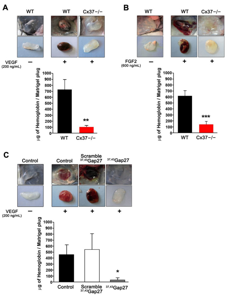
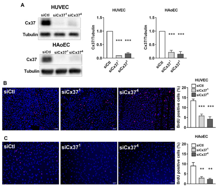
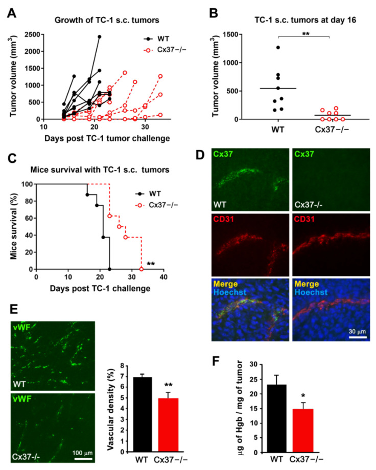
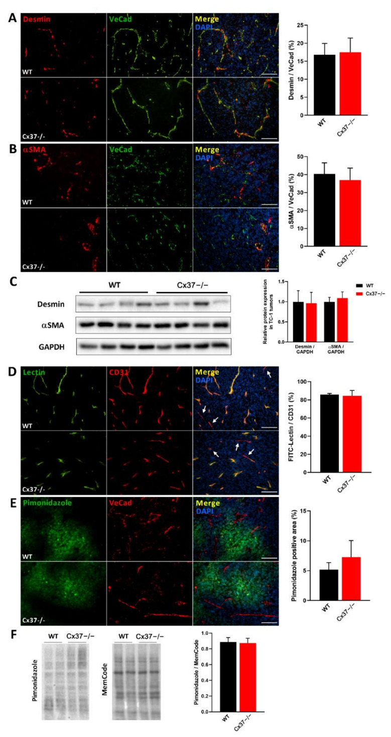
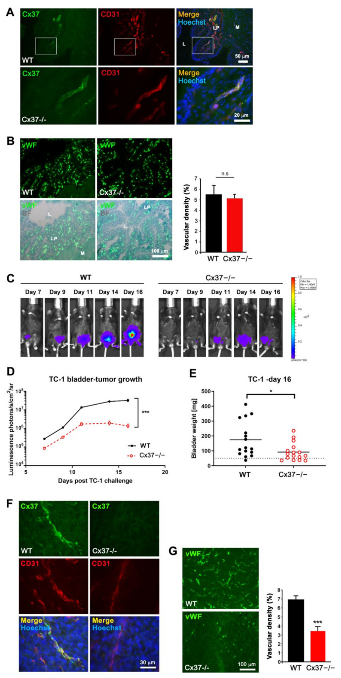
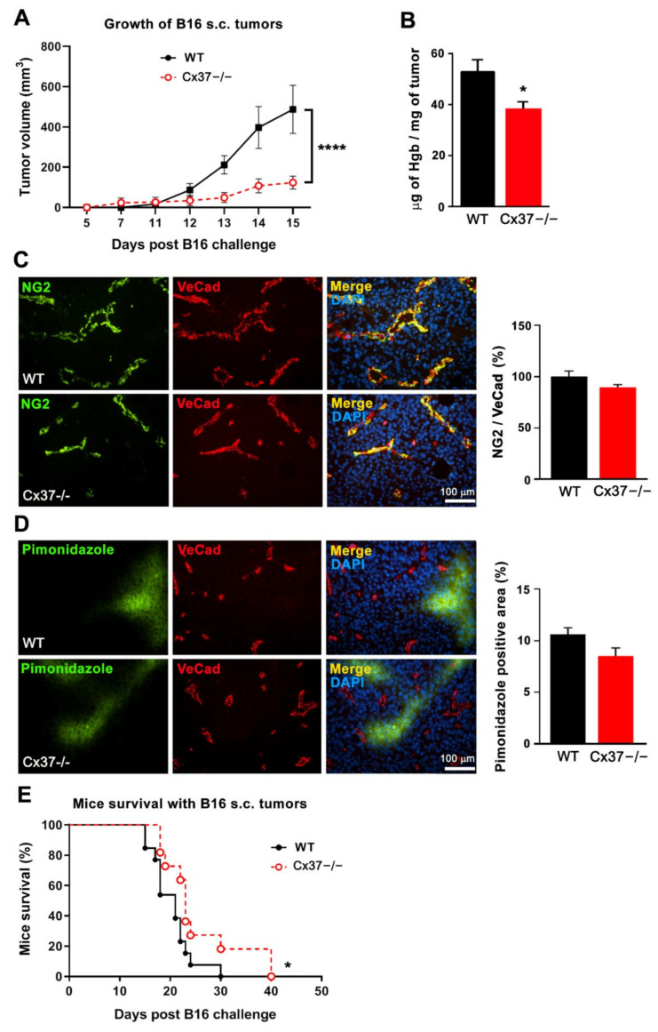
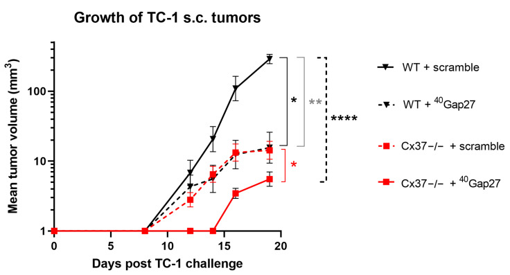
Connexin37 (Cx37) and Cx40 form intercellular channels between endothelial cells (EC), which contribute to the regulation of the functions of vessels. We previously documented the participation of both Cx in developmental angiogenesis and have further shown that loss of Cx40 decreases the growth of different tumors. Here, we report that loss of Cx37 reduces (1) the in vitro proliferation of primary human EC; (2) the vascularization of subcutaneously implanted matrigel plugs in Cx37-/- mice or in WT using matrigel plugs supplemented with a peptide targeting Cx37 channels; (3) tumor angiogenesis; and (4) the growth of TC-1 and B16 tumors, resulting in a longer mice survival. We further document that Cx37 and Cx40 function in a collaborative manner to promote tumor growth, inasmuch as the injection of a peptide targeting Cx40 into Cx37-/- mice decreased the growth of TC-1 tumors to a larger extent than after loss of Cx37. This loss did not alter vessel perfusion, mural cells coverage and tumor hypoxia compared to tumors grown in WT mice. The data show that Cx37 is relevant for the control of EC proliferation and growth in different tumor models, suggesting that it may be a target, alone or in combination with Cx40, in the development of anti-tumoral treatments.

Keywords: angiogenesis; cell-cell communication; connexins; transgenic mice; tumors.


Conflict of interest statement

The authors declare no conflict of interest. The funders had no role in the design of the study; in the collection, analyses or interpretation of data; in the writing of the manuscript; or in the decision to publish the results.

Figures

Figure 1
Figure 1
Loss of Cx37 reduces angiogenesis in subcutaneous matrigel plugs. (A,B) Representative pictures of matrigel plugs without and with VEGF (A) or FGF2 (B), one week after s.c. injection in WT and Cx37−/− mice. In the absence of vascular growth factors, the plugs retained their original whitish color, due to the absence of vascularization. In the presence of the growth factors, the plugs implanted in WT mice acquired a red-brown color, reflecting their neovascularization, as also judged by a sizable hemoglobin content. Both the color change and the hemoglobin content of the plugs were significantly lower in the plugs implanted in Cx37−/− mice. Data are means + SEM. n = 13 WT and 9 Cx37−/− mice. ** p < 0.01, *** p < 0.001 versus WT mice (Student’s t-test). (C) Matrigel plugs containing VEGF were supplemented with either the 37,43Gap27 peptide or its scrambled version, before implantation in WT mice. The peptide targeting Cx37 markedly reduced the color change and the hemoglobin content of the plugs, which retained the whitish appearance of the controls devoid of VEGF, and which was similar to that observed in Cx37−/− mice. In contrast, plugs loaded with VEGF and the scrambled peptide became red-stained and featured a large increase in hemoglobin content, as observed in WT mice. Data are mean + SEM. n = 5–6 mice per group. * p < 0.05 versus scramble (one-way ANOVA followed by Tukey’s post-test).
Figure 2
Figure 2
In vitro targeting Cx37 reduces the proliferation of primary EC. (A) Western blot of primary human vein (HUVEC) and aortic EC (HAoEC) revealed that Cx37 expression was reduced by 80–90% 48 h after transfection with either siCx371 or siCx374, two siRNAs targeting Cx37. Such a drastic change was not observed when the cells were transfected with a control siRNA (siCtl). Graphs show mean + SEM, *** p < 0.001 versus siCtl (one-way ANOVA followed by Tukey’s post-test). (B,C) As judged by the incorporation of BrdU (red) in the nuclei (stained in blue by DAPI), the proliferation of the HUVEC and HAoEC that had lost most of Cx37 was decreased to about 30% of control levels. Graphs show mean values + SEM, n = 8 separate experiments, each performed in duplicate. ** p < 0.01, *** p < 0.001 vs. siCtl (one-way ANOVA followed by Tukey’s post-test). Scale bars = 50 μm.
Figure 3
Figure 3
The growth of TC-1 tumors is reduced in Cx37−/− mice. (A) After s.c. injection, TC-1 cells generated growing tumors in all WT and Cx37−/− mice. (B) Sixteen days after the cell injection, the volume of tumors was significantly lower in Cx37−/− (n = 8) than in WT mice (n = 8). ** p < 0.01 versus WT mice (Student’s t-test). The horizontal bars show the mean tumor volume in each group. (C) All WT mice carrying the TC-1 tumor were sacrificed within the first 3 weeks of the experiments. In contrast, the cognate Cx37−/− mice survived significantly longer. ** p = 0.01 versus WT mice (log-rank Mantel-Cox test). (D) Sixteen days after the injection of TC-1 cells, Cx37 was detected via immunostaining in CD31-positive EC of the tumors grown in WT (left) but not Cx37−/− mice (right). Bar, 30 µm. (E) Immunostaining of the EC-specific von Willebrand factor (vWF) revealed a lower density of vessels in the tumors grown in Cx37−/− than in WT mice. Data are mean + SEM of 5–7 areas from size-matched tumors that developed in 4 mice per group. ** p < 0.01 versus WT mice (Student’s t-test). Bar, 100 µm. (F) The hemoglobin concentration was also lower in the tumors that developed in Cx37−/− than WT mice. Data are mean + SEM. n = 6 mice per group. * p < 0.05 vs. WT mice (Student’s t-test).
Figure 4
Figure 4
Loss of Cx37 does not alter the maturation of vessels in TC-1 tumors. (A,B) Left panels: immunostaining of desmin (A) and alpha-smooth muscle actin (αSMA; (B)) shows the mural cells which surrounded VeCad-positive EC in the newly formed vessels of TC-1 tumors. Right panels: The right bar graphs show that the coverage of vessels by mural cells was similar in the tumors grown in Cx37−/− and WT mice. Data are mean + SEM of 8–15 fields, from at least 5 mice per group. Bars = 100 μm. (C) Western blots confirmed that the tumors grown in Cx37−/− and WT mice featured similar levels of desmin and αSMA. Data are mean + SEM. n = 7 WT and 8 Cx37−/− mice (Student’s t-test). (D) Infusion of FITC-labeled tomato lectin (green) stained most CD31-positive vessels (red) of TC-1 tumors in both Cx37−/− and WT mice (white arrows indicate some of the few vessels that were not perfused by the lectin). Quantitative analysis showed that a similar proportion of tumor vessels was perfused in the two types of animals. Data are mean + SEM of 6–8 fields from 4 mice per group (Student’s t-test). (E) Immunostaining of pimonidazole (green) revealed hypoxic areas in tumor regions containing VeCad-positive vessels (red). Quantitative analysis revealed that the percentage of these hypoxic areas was comparable in the tumors grown in Cx37−/− and WT mice. Data are mean + SEM of 8–15 fields from 7 mice per group (Student’s t-test). Bars = 100 μm. (F) Western blots of total proteins extracted from TC-1 tumors confirmed comparable levels of pimonidazole-protein adducts in the tumors grown in Cx37−/− and WT mice. Total protein stain (MemCode) was used to control for a comparable protein loading. The specificity of the pimonidazole staining was assessed using tumor samples of WT and Cx37−/− mice which had not been previously injected with the pimonidazole HCl (HypoxyprobeTM) and revealed no adducts’ signal (data not shown). Data are mean + SEM. WT (n = 8) and Cx37−/− (n = 7) mice (Student’s t-test).
Figure 5
Figure 5
Loss of Cx37 decreases the vascularization and growth of TC-1 tumors established in the bladder. (A) Immunostaining showed Cx37 (green) in the CD31-positive EC (red) of control bladders. Representative images are shown at low (bar = 50 µm) and high magnification (bar = 20 µm) in the top and bottom row, respectively. Cell nuclei are seen after Hoechst staining (blue). (B) Immunostaining of native bladder sections for Von Willebrand factor (vWF, top panel), overlaid with Evan’s blue counterstain (bright field, BF, bottom panel), revealed many blood vessels, mainly localized within the lamina propria of the bladder (LP; L= lumen; M: muscular layers). Quantitative evaluation showed that the volume density of these vessels was similar in the bladders of WT and Cx37−/− mice. Data are mean + SEM values of 3 fields, photographed from 3 mice per group. (C) Representative bioluminescence imaging of TC-1-luc tumors growing in the bladder of a WT (left) and a Cx37−/− mouse (right). (D) The mean + SEM bioluminescence intensity (photons/sec/cm2/sr) of the TC-1 tumors growing in the bladder of WT mice (black line, n = 16) was significantly higher than that of the tumors growing in the bladder of Cx37−/− mice (red line, n = 16). *** p < 0.001 versus WT mice (Student’s t-test on the area under the tumor growth curve). (E) Sixteen days after the intravesical instillation of TC-1 cells, the combined weight of bladders and growing tumors was measured and found to be significantly higher in WT than Cx37−/− mice. Horizontal lines show mean values. The dotted line shows the mean weight of native mice bladders. * p < 0.05 versus WT mice (Student’s t-test). (F) Immunostaining showed the presence of Cx37 on CD31-positive EC of TC-1-luc tumors grown in the bladders of WT (left) but not of Cx37−/− mice (right). (G) After 16 days, the immunostaining for von Willebrand factor (vWF) revealed that the vessel density of TC-1-luc tumors was higher in Cx37−/− than WT mice. Data are mean + SEM of 5–7 areas from tumors of similar size, which developed in 5 different mice per group. *** p < 0.001 versus WT mice (Student’s t-test).
Figure 6
Figure 6
Loss of Connexin37 decreases the growth of B16-F10 tumors and extends the survival of the tumor-bearing mice. B16-F10 cells were s.c. injected to generate tumors in WT and Cx37−/− mice. (A) The volume of the tumors growing in Cx37−/− mice until day 15 was significantly smaller than that of the tumors growing in WT animals. Data are mean ± SEM of WT mice (n = 11, black line) and Cx37−/− mice (n = 13, dotted red line). **** p< 0.0001 versus WT mice (areas under the tumor growth curve were compared by Student’s t-test). (B) In another set of mice, which were sacrificed 14 days after the cell injection, the hemoglobin content of the tumors grown in Cx37−/− mice was lower than that evaluated in the WT controls. (C) Immunostaining showed a similar distribution of NG2 over the VeCad positive vessels of the B16-F10 tumors which grew in WT and Cx37−/− mice. (D) Immunostaining of pimonidazole (green) revealed hypoxic areas in tumor regions containing VeCad-positive vessels (red). Quantitative analysis revealed that the surface of these hypoxic areas was comparable in the tumors grown in Cx37−/− and WT mice. Data are mean + SEM of 8–15 fields from 7 mice per group (Student’s t-test). Bars = 100 μm. (E) The survival of the Cx37−/− mice that carried a B16 tumor was significantly increased compared to that of the cognate WT animals. Data are mean ± SEM of 11 WT mice (black line) and 13 Cx37−/− mice (dotted red line). * p < 0.05 versus WT mice (log-rank Mantel-Cox test).
Figure 7
Figure 7
A peptide targeting Cx40 function decreases tumoral growth in Cx37−/− mice. One day after the s.c. injection of TC-1 cells, WT (n = 12, black lines) and Cx37−/− mice (n = 16, red lines) received a daily i.p. injection of either the 40Gap27 peptide, which specifically targets Cx40 channels, or its scrambled version, which served as control. The plot shows the mean ± SEM tumor volumes as a function of time. After 19 days, the growth of TC-1 tumors that received the scramble version of the peptide was significantly slower in Cx37−/− than in WT mice. In both groups of mice, the animals that received the 40Gap27 peptide developed smaller tumors than the animals which received its scrambled form. Significant differences in tumor growth, as judged by the area under the curve, were determined using a one-way ANOVA and a Sidak’s multiple comparisons post-test. Cx37−/− + scramble vs. WT + scramble (grey); Cx37−/− + scramble vs. Cx37−/− + 40Gap27 (red); WT + 40Gap27 vs. WT+ scramble (black); Cx37−/− + 40Gap27 vs. WT+ scramble (black dash line). * p < 0.05; ** p < 0.01; **** p < 0.0001.

Similar articles

Cited by

References

    1. Li X., Li Y., Lu W., Chen M., Ye W., Zhang D. The Tumor Vessel Targeting Strategy: A Double-Edged Sword in Tumor Metastasis. Cells. 2019;8:1602. doi: 10.3390/cells8121602. - DOI - PMC - PubMed
    1. Chung A.S., Ferrara N. Developmental and pathological angiogenesis. Annu. Rev. Cell Dev. Biol. 2011;27:563–584. doi: 10.1146/annurev-cellbio-092910-154002. - DOI - PubMed
    1. Goel S., Duda D.G., Xu L., Munn L.L., Boucher Y., Fukumura D., Jain R.K. Normalization of the vasculature for treatment of cancer and other diseases. Physiol. Rev. 2011;91:1071–1121. doi: 10.1152/physrev.00038.2010. - DOI - PMC - PubMed
    1. Tahergorabi Z., Khazaei M. Imbalance of angiogenesis in diabetic complications: The mechanisms. Int. J. Prev. Med. 2012;3:827–838. doi: 10.4103/2008-7802.104853. - DOI - PMC - PubMed
    1. Nohata N., Uchida Y., Stratman A.N., Adams R.H., Zheng Y., Weinstein B.M., Mukouyama Y.-S., Gutkind J.S. Temporal-specific roles of Rac1 during vascular development and retinal angiogenesis. Dev. Biol. 2016;411:183–194. doi: 10.1016/j.ydbio.2016.02.005. - DOI - PubMed

LinkOut - more resources