. 2022 Mar 9;23(6):2948.
doi: 10.3390/ijms23062948.

Therapeutic Effects of Platelet-Derived Extracellular Vesicles in a Bioengineered Tendon Disease Model

Affiliations

Therapeutic Effects of Platelet-Derived Extracellular Vesicles in a Bioengineered Tendon Disease Model

Ana L Graça et al. Int J Mol Sci. .

Abstract

Tendon injuries represent over 30-50% of musculoskeletal disorders worldwide, yet the available therapies do not provide complete tendon repair/regeneration and full functionality restoring. Extracellular vesicles (EVs), membrane-enclosed nanoparticles, have emerged as the next breakthrough in tissue engineering and regenerative medicine to promote endogenous tissue regeneration. Here, we developed a 3D human in vitro model mimicking the signature of pathological tendon and used it to evaluate the influence that different platelet-derived EVs might have in tendon tissue repair mechanisms. For this, different EV populations isolated from platelets, small EVs (sEVs) and medium EVs (mEVs), were added to the culture media of human tendon-derived cells (hTDCs) cultured on isotropic nanofibrous scaffolds. The platelet-derived EVs increased the expression of tenogenic markers, promoted a healthy extracellular matrix (ECM) remodeling, and the synthesis of anti-inflammatory mediators. These findings suggest that platelet EVs provided relevant biochemical cues that potentiated a recovery of hTDCs phenotype from a diseased to a healthy state. Thus, this study opens new perspectives for the translation of platelet-derived EVs as therapeutics.

Keywords: extracellular vesicles; fibers; in vitro models; platelets; tendinopathy; tendon-derived cells; tissue engineering.


Conflict of interest statement

The authors declare no conflict of interest.

Figures

Figure 1
Figure 1
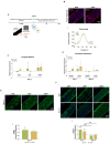
(A) (i) Scanning electron microscopy (SEM) micrographs of the produced misaligned fiber threads. Scale bars: 100 µm and 20 µm. (ii) Isotropic fibers and threads diameter. Data are presented as mean ± standard deviation (SD) (n = 50). (iii) Platelet lysate (PL) coating at day 0. Scale bar: 200 µm. (B) (i) SEM images of small and medium extracellular vesicles (sEVs; mEVs) isolated from PL. Scale bar: 300 nm. (ii) Size distribution profile of sEVs and mEVs analyzed with nanoparticle tracking analysis (NTA) (n = 3). (iii) EV basic fibroblast growth factor (bFGF), transforming growth factor-beta 1 (TGF-β1), vascular endothelial growth factor-A (VEGF-A), and platelet-derived growth factor-BB (PDGF-BB) quantification. Data are presented as mean ± SD (n = 3). Statistical significance: * p < 0.05, ** p < 0.01.
Figure 2
Figure 2
(A) Schematic representation of human tendon-derived stem cells (hTDCs) in culture over time. hTDCs were maintained in culture for 14 days to acquire a disease phenotype. Afterward, small extracellular vesicles (sEVs) and medium extracellular vesicles (mEVs) were added to the culture media of hTDCs to recover their healthy phenotype, which was evaluated after 7 and 14 days of culture. (B) (i) Confocal microscopy images of F-actin filaments (DAPI, blue; Phalloidin, red) of hTDCs supplemented with sEVs and mEVs after 14 days of culture. Scale bar: 75 µm. (ii) Representative directionality analysis of F-actin filaments in hTDCs after 14 days (n = 3). (C) Gene expression profile of tenogenic markers (mohawk (MKX), scleraxis (SCX), tenomodulin (TNMD)), after 7 days of culture (n = 4). Gene expression results are presented as fold changes with respect to the control group at each time point (represented by a line at 1). Statistical differences: ° p < 0.05 (against to the control). (D) (i) Confocal images of TNMD (green) at day 14. Images were counterstained with cells nuclei (DAPI, blue). Scale bar: 75 µm. (ii) TNMD fluorescence intensity quantitative analysis. Data were normalized to the control (1). Data are presented as mean ± standard deviation (SD) (n = 6). Statistical differences: ° p < 0.05 (against to the control). (E) Gene expression profile of phenotypic drift markers (runt-related transcription factor 2 (RUNX2) and SRY-Box transcription factor (SOX9)), after 7 and 14 days of culture. Gene expression results are presented as fold changes with respect to the control group at each time point (represented by a line at 1) (n = 4). (F) (i) Confocal micrographs of alpha-smooth muscle actin (ACTA2; green) at 7 and 14 days of culture. Nuclei were counterstained with DAPI (blue). Scale bar: 75 µm. (ii) Quantification of ACTA2 fluorescence intensity. Data were normalized to the control (1). Data are presented as mean ± SD (n = 6). Statistical differences: *** p < 0.001, **** p < 0.0001 (among days for the same group); °° p < 0.01, °°°° p < 0.0001 (against to the control).
Figure 3
Figure 3
(A) Gene expression of tendon-related extracellular matrix (ECM) markers, collagen type I alpha 1 chain (COL1A1), collagen type III alpha 1 chain (COL3A1), decorin (DCN), and tenascin (TNC), after 7 and 14 days with small extracellular vesicles (sEVs) and medium extracellular vesicles (mEVs). Expression of target genes normalized against the reference genes glyceraldehyde-3-phosphate dehydrogenase (GADPH) and glucuronidase beta (GUSB) and gene expression normalized at the control of each day (1) (n = 4). Statistical differences: * p < 0.05, ** p < 0.01 (among days for the same group); ° p < 0.05 (sEVs against mEVs). (B) Concentration of matrix metalloproteinase-1, -2, -3, -7 and -9 (MMP-1, MMP-2, MMP-3, MMP-7, and MMP-9) and tissue inhibitor of metalloproteinase-1 (TIMP-1), present in the 14 days culture medium. Data were normalized to the control of day 14 (unstimulated cells, set to 1) (n = 4). Statistical differences: ° p < 0.05 (sEVs against mEVs). (C) Venn diagram of common and unique ECM proteins identified in the isotropic fiber-encapsulated human tendon-derived stem cell (hTDC) system, non-supplemented and supplemented with sEVs and mEVs at 14 days of culture (n = 3). (D) Relative abundance of ECM proteins was categorized according to their function. Quantitative assessment of the ECM proteomic profile revealed (i) matricellular and (ii) fibrillar collagen are the most abundant categories (n = 3). Statistical differences: °°°° p < 0.0001 (against control), **** p < 0.0001 (sEVs against mEVs) (E) Proteomic profile assessment of collagen type I and collagen type III (COL1/COL3) ratio at 14 days of culture (n = 3). Results were normalized to control that was set to 1.
Figure 4
Figure 4
Gene expression of inflammatory markers (interleukin-4; -6; -8 (IL4, IL6, and IL8)), after 7 and 14 days of culture with EVs. The target gene expression was normalized against glyceraldehyde-3-phosphate dehydrogenase (GAPDH) and glucuronidase beta (GUSB) and the data presented for the non-supplemented system each day (1) (n = 4). Statistical significances: * p < 0.05 (among days for the same group); ° p < 0.05 (between sEVs and mEVs or against the control for the same day).

Similar articles

Cited by

References

    1. Millar N.L., Silbernagel K.G., Thorborg K., Kirwan P.D., Galatz L.M., Abrams G.D., Murrell G.A.C., McInnes I.B., Rodeo S.A. Tendinopathy. Nat. Rev. Dis. Prim. 2021;7:1–21. doi: 10.1038/s41572-020-00234-1. - DOI - PubMed
    1. Millar N.L., Murrell G.A.C., McInnes I.B. Inflammatory mechanisms in tendinopathy—Towards translation. Nat. Rev. Rheumatol. 2017;13:110–122. doi: 10.1038/nrrheum.2016.213. - DOI - PubMed
    1. Leadbetter W.B. Anti-inflammatory therapy in tendinopathy: The role of nonsteroidal drugs and corticosteroid injections. Tendon Inj. Basic Sci. Clin. Med. 2005:211–232. doi: 10.1007/1-84628-050-8_22. - DOI - PubMed
    1. Aicale R., Bisaccia R.D., Oliviero A., Oliva F., Maffulli N. Current pharmacological approaches to the treatment of tendinopathy. Expert Opin. Pharmacother. 2020;21:1467–1477. doi: 10.1080/14656566.2020.1763306. - DOI - PubMed
    1. Mishra A., Woodall J., Vieira A. Treatment of Tendon and Muscle Using Platelet-Rich Plasma. Clin. Sports Med. 2009;28:113–125. doi: 10.1016/j.csm.2008.08.007. - DOI - PubMed

LinkOut - more resources