. 2022 Mar 9;23(6):2964.
doi: 10.3390/ijms23062964.

The Combination of Intestinal Alkaline Phosphatase Treatment with Moderate Physical Activity Alleviates the Severity of Experimental Colitis in Obese Mice via Modulation of Gut Microbiota, Attenuation of Proinflammatory Cytokines, Oxidative Stress Biomarkers and DNA Oxidative Damage in Colonic Mucosa

Affiliations

The Combination of Intestinal Alkaline Phosphatase Treatment with Moderate Physical Activity Alleviates the Severity of Experimental Colitis in Obese Mice via Modulation of Gut Microbiota, Attenuation of Proinflammatory Cytokines, Oxidative Stress Biomarkers and DNA Oxidative Damage in Colonic Mucosa

Dagmara Wojcik-Grzybek et al. Int J Mol Sci. .

Abstract

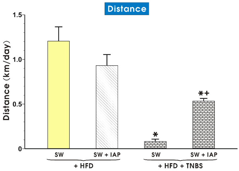
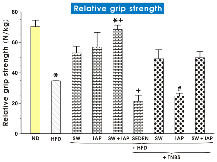
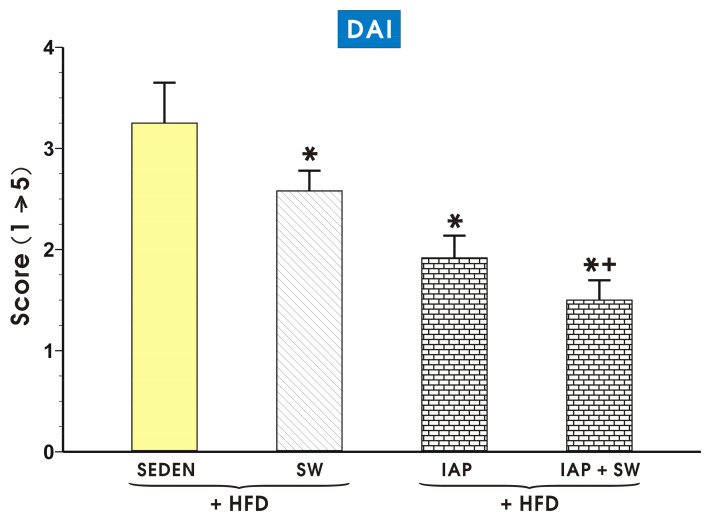
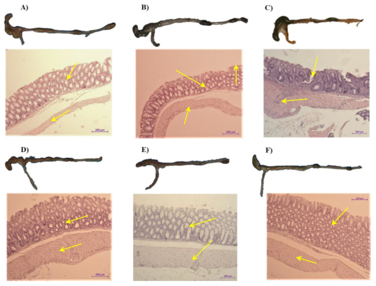
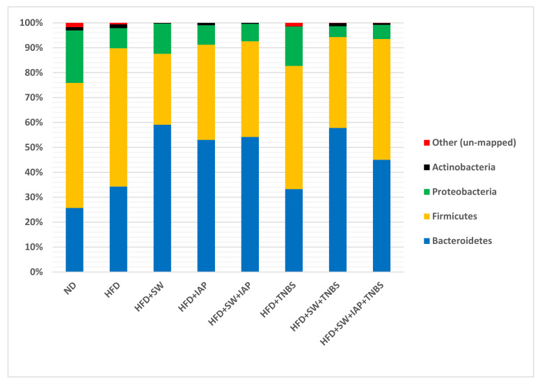
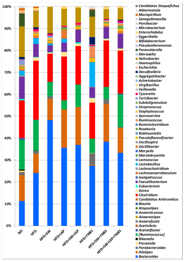
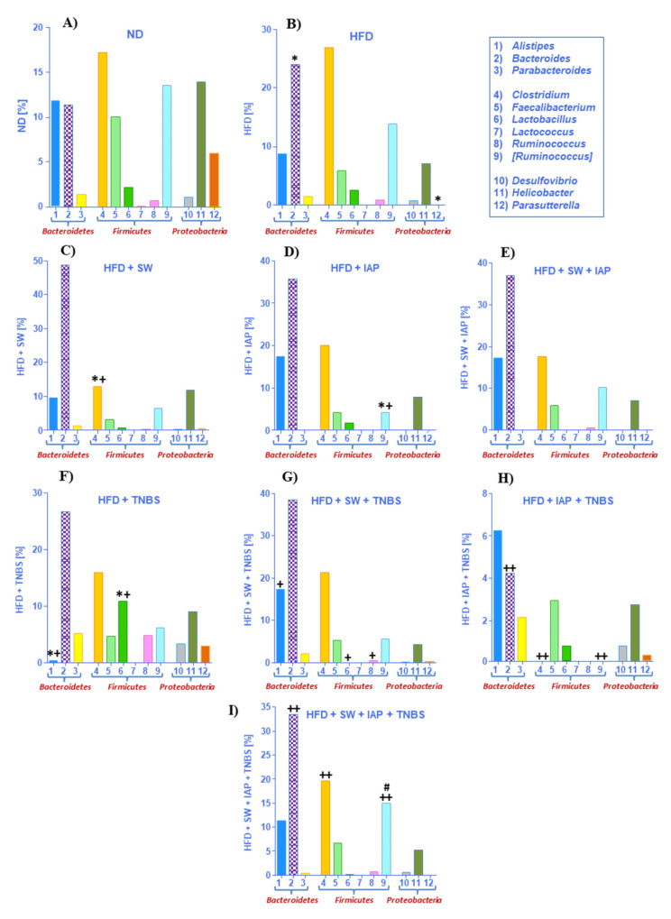
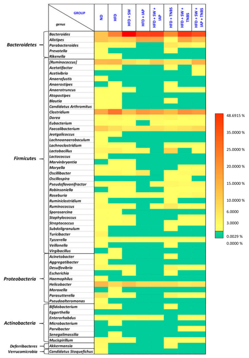
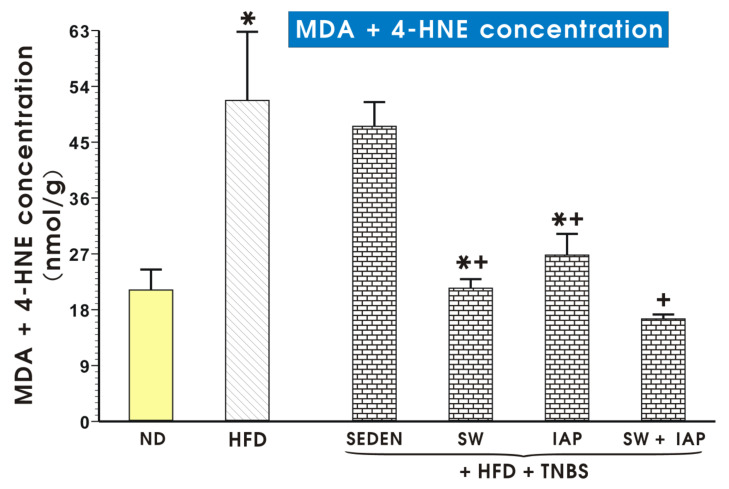
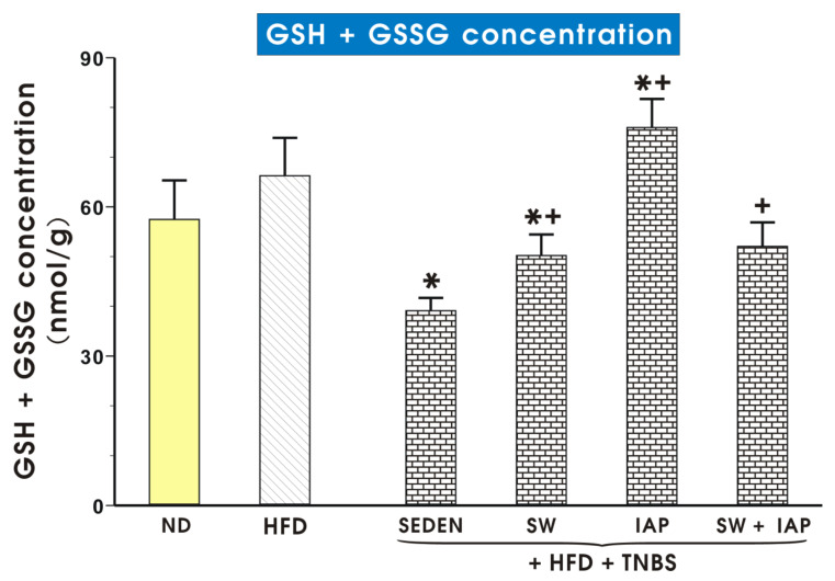
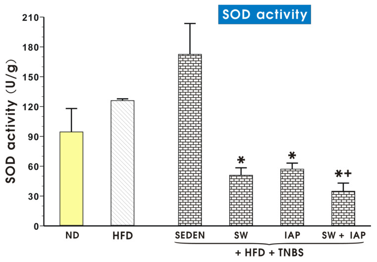
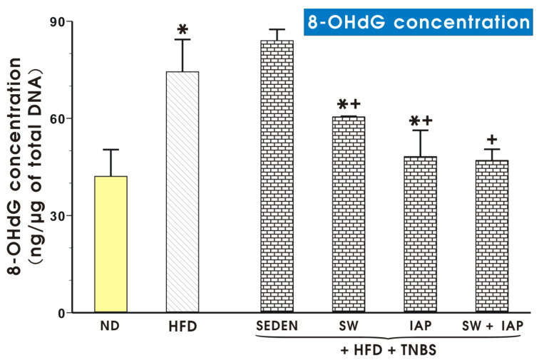
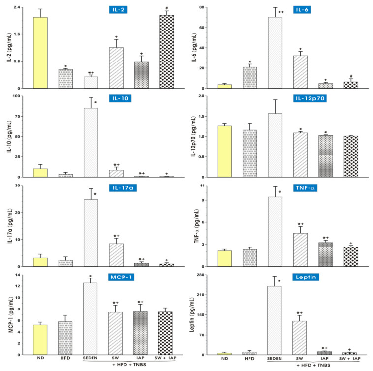
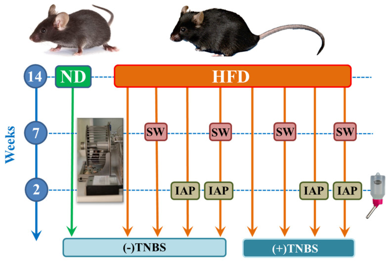
Inflammatory bowel diseases (IBD) are commonly considered as Crohn's disease and ulcerative colitis, but the possibility that the alterations in gut microbiota and oxidative stress may affect the course of experimental colitis in obese physically exercising mice treated with the intestinal alkaline phosphatase (IAP) has been little elucidated. Mice fed a high-fat-diet (HFD) or normal diet (ND) for 14 weeks were randomly assigned to exercise on spinning wheels (SW) for 7 weeks and treated with IAP followed by intrarectal administration of TNBS. The disease activity index (DAI), grip muscle strength test, oxidative stress biomarkers (MDA, SOD, GSH), DNA damage (8-OHdG), the plasma levels of cytokines IL-2, IL-6, IL-10, IL-12p70, IL-17a, TNF-α, MCP-1 and leptin were assessed, and the stool composition of the intestinal microbiota was determined by next generation sequencing (NGS). The TNBS-induced colitis was worsened in obese sedentary mice as manifested by severe colonic damage, an increase in DAI, oxidative stress biomarkers, DNA damage and decreased muscle strength. The longer running distance and weight loss was observed in mice given IAP or subjected to IAP + SW compared to sedentary ones. Less heterogeneous microbial composition was noticed in sedentary obese colitis mice and this effect disappeared in IAP + SW mice. Absence of Alistipes, lower proportion of Turicibacter, Proteobacteria and Faecalibacterium, an increase in Firmicutes and Clostridium, a decrease in oxidative stress biomarkers, 8-OHdG content and proinflammatory cytokines were observed in IAP + SW mice. IAP supplementation in combination with moderate physical activity attenuates the severity of murine colitis complicated by obesity through a mechanism involving the downregulation of the intestinal cytokine/chemokine network and oxidative stress, the modulation of the gut microbiota and an improvement of muscle strength.

Keywords: diet-induced obesity; experimental colitis; inflammation; intestinal alkaline phosphatase; oxidative stress; voluntary exercise.


Conflict of interest statement

The authors declare no conflict of interest.

Figures

Figure 1
Figure 1
The running distance in obese exercising mice subjected to voluntary exercise on spinning wheels (SW) alone, SW combined or not combined with intestinal alkaline phosphatase (IAP) administration with or without TNBS colitis. Results are mean ± S.E.M. of 6–8 animals. An asterisk indicates a significant change (p < 0.05) as compared to the respective values in exercising mice fed a high fat diet (HFD). An asterisk and cross indicate a significant change (p < 0.05) as compared to the values in exercising obese mice with or without colitis.
Figure 2
Figure 2
Relative grip strength in sedentary mice fed a normal diet (ND) or high fat diet (HFD) with or without TNBS colitis subjected to voluntary exercise on the spinning wheels (SW) or a treatment with the intestinal alkaline phosphatase (IAP) alone or combined with SW (SW + IAP). Results are mean ± S.E.M. of 7 animals per group. An asterisk indicates a significant change (p < 0.05) as compared to the respective values in mice fed ND. An asterisk with cross indicates a significant change (p < 0.05) as compared to the respective values in obese mice subjected to SW or treated with IAP alone. A cross indicates a significant change (p < 0.05) as compared to the respective values in HFD fed sedentary (SEDEN) mice. A hash indicates a significant decrease (p < 0.05) as compared to the respective values obtained in obese TNBS mice subjected to SW only or the combination of SW + IAP.
Figure 3
Figure 3
The effect of exercise on spinning wheels (SW) and intestinal alkaline phosphatase (IAP) administration both applied alone or in the combination SW+ IAP on the disease activity index (DAI) in obese mice (HFD fed) with TNBS-induced colitis as compared to sedentary (SEDEN) obese mice with TNBS colitis. Results are mean ± S.E.M. of 8 animals per each group. An asterisk indicates a significant change (p < 0.05) as compared to the respective value in the obese SEDEN mice with colitis. An asterisk and cross indicate a significant change (p < 0.05) as compared to the respective values in the obese mice with TNBS-induced colitis subjected to SW only or to the treatment with IAP alone.
Figure 4
Figure 4
The representative macroscopic and microscopic appearance of the colon obtained from: (A) a sedentary mouse without colitis and fed a normal diet (ND); (B) a sedentary mouse without colitis and fed a high fat diet (HFD); (C) an obese mouse fed HFD with TNBS-induced colitis; (D) an obese mouse with TNBS colitis, subjected to exercise on spinning wheels (SW); (E) an obese mouse with the intestinal alkaline phosphatase (IAP) administration with TNBS colitis; (F) an obese mouse with TNBS colitis and subjected to the combination of exercise on SW and administered with IAP. The histology photomicrographs represent: (A) a colon from a sedentary mouse without colitis and fed a ND: normal macroscopic appearance of the colonic mucosa and no histologic injury to mucosa and submucosa as indicated by arrows; (B) a colon from a sedentary mouse fed a HFD: normal macroscopic appearance of the colonic mucosa and no histologic injury to mucosa and submucosa as indicated by arrows, (C) a colon from an obese, sedentary mouse with colitis: intestinal damage, hemorrhagic lesions, prominent mucosal inflammation and a bloody effusion of colonic mucosa and submucosa (arrows); (D) a colon from an obese mouse with voluntary exercise on SW and TNBS-induced colitis: fewer hemorrhagic lesions and mild inflammatory reaction in submucosa (arrows); (E) a colon from an obese mouse with IAP administration and TNBS-induced colitis: less inflammatory reaction in mucosa and submucosa, signs of regeneration of hypertrophic mucosa (arrows); (F) a colon from an obese mouse subjected to the combination of exercise on SW and IAP treatment with TNBS-induced colitis: marked improvement in histopathology of colonic mucosa as manifested by a preservation of mucosal lining, hypertrophic epithelium and a reduction in bleeding and hemorrhagic lesions (arrows).
Figure 5
Figure 5
Relative abundance (%) of the gut microbiome at the phylum level for mice fed a normal diet (ND) only (control) or a high fat diet (HFD) with or without TNBS colitis administered with intestinal alkaline phosphatase (IAP) and subjected or not to voluntary exercise on spinning wheels (SW).
Figure 6
Figure 6
Relative abundance (%) of the gut microbiome at the genera level for mice fed a normal diet (ND) only or fed a high fat diet (HFD) with or without TNBS-induced colitis administered with intestinal alkaline phosphatase (IAP) alone, or exercising on spinning wheels (SW) only or subjected to the combination of SW and IAP (SW + IAP).
Figure 7
Figure 7
Relative abundance of the gut microbiome for the most important genus of mice fed normal diet (ND) (A) or high fat diet (HFD) (B) only, obese mice with spinning wheel (SW) exercise (HFD + SW) (C), obese mice administered with intestinal alkaline phosphatase (IAP) alone (HFD + IAP) (D) or subjected to the combination of SW and IAP without TNBS colitis (HFD + SW + IAP) (E) and obese mice with TNBS colitis (HFD + TNBS) (F), obese mice with TNBS colitis exercising on SW (HFD + SW + TNBS) (G), obese mice with TNBS colitis administered with IAP alone (HFD + IAP + TNBS) (H) or obese mice administered with IAP in combination with SW (HFD + SW + IAP + TNBS) (I). An asterisk indicates a significant change (p < 0.05) as compared to the respective values in the sedentary mice fed ND. An asterisk and cross indicate a significant change (p < 0.05) as compared to the respective values in the sedentary mice fed a HFD. A cross indicates a significant change (p < 0.05) as compared to the respective values in the HFD fed mice with TNBS colitis. A double cross indicates a significant change (p < 0.05) as compared to the respective values in HFD fed mice with or without access to SW with TNBS colitis. Hash indicates a significant change (p < 0.05) as compared to the respective values in HFD fed TNBS colitis mice with SW only or treatment with IAP alone.
Figure 8
Figure 8
Heat map of species abundance clustering at the genus level, estimated from 16S rRNA amplicon sequencing, of gut microbiome of mice in mice fed a normal diet (ND) or high fat diet (HFD) alone, in mice fed a HFD and subjected to voluntary exercise on spinning wheels (SW) (HFD + SW), mice fed a HFD and treated with the intestinal alkaline phosphatase (IAP) (HFD + IAP), mice fed a HFD with TNBS colitis (HFD + TNBS), mice fed HFD with IAP administration (HFD + SW + IAP), mice fed HFD with access to SW with TNBS colitis (HFD + SW + TNBS) and in TNBS colitis mice fed HFD with the combination of SW and IAP administration (HFD + SW + IAP + TNBS). Color reflects relative abundance from low (yellow) to high (red) with green as zero.
Figure 9
Figure 9
Lipid peroxidation products expressed as malondialdehyde and 4-hydroxynonenal (MDA + 4-HNE) concentration in colonic mucosa of a normal diet (ND) or obese mice, voluntary exercised TNBS colitis mice exercising on spinning wheels (SW) with or without the intestinal phosphatase (IAP) treatment or in those subjected to the combination of SW and IAP. Results are mean ± S.E.M. of 6–8 animals per group. An asterisk indicates a significant change (p < 0.05) as compared to the respective values in the sedentary mice (SEDEN) fed ND. An asterisk with cross indicates a significant change (p < 0.05) as compared to the respective values in the sedentary mice fed a HFD with induced colitis. A cross indicates a significant change as compared to the respective values in the HFD mice with administration of IAP and induced colitis (p < 0.05).
Figure 10
Figure 10
Total glutathione (reduced GSH+ oxidized GSSG) concentration in colonic mucosa in mice with or without TNBS colitis fed a normal diet (ND) or a high fat diet (HFD) with access to spinning wheels (SW), administered with or without combination with the intestinal alkaline phosphatase (IAP). Results are mean ± S.E.M. of 6–8 animals per each group. An asterisk indicates a significant change (p < 0.05) as compared to the respective values in the sedentary mice (SEDEN) fed HFD. An asterisk with cross indicates a significant change (p < 0.05) as compared to the respective values in the sedentary TNBS colitis mice fed a HFD. A cross indicates a significant change as compared to the respective values in obese TNBS mice treated with IAP (p < 0.05).
Figure 11
Figure 11
Determination superoxide dismutase (SOD) activity in colonic mucosa of a normal diet (ND) or obese mice fed a high fat diet (HFD) with or without TNBS colitis with access to spinning wheels (SW) with or without the intestinal alkaline phosphatase (IAP) treatment. Results are mean ± S.E.M. of 6–8 animals per group. An asterisk indicates a significant change (p < 0.05) as compared to the respective values in sedentary (SEDEN) mice fed a HFD. An asterisk with a cross indicates a significant change (p < 0.05) as compared to the respective values in the HFD fed mice with colitis subjected to SW only or treated with IAP alone.
Figure 12
Figure 12
DNA oxidative damage in colonic mucosa of a normal diet (ND) or a high fat diet (HFD) mice with or without TNBS colitis subjected to exercise on spinning wheels (SW) or the intestinal alkaline phosphatase (IAP) treatment alone or the combination of SW and IAP. Results are mean ± S.E.M. of 6–8 animals per group. An asterisk indicates a significant change (p < 0.05) as compared to the respective values in the control mice fed an ND. An asterisk with cross indicates a significant change (p < 0.05) as compared to the respective values in the sedentary mice (SEDEN) with TNBS-induced colitis fed a HFD. A cross indicates a significant change (p < 0.05) as compared to the respective values in the HFD fed colitis mice with access to SW.
Figure 13
Figure 13
Plasma level of interleukin-2 (IL-2), IL-6, IL-10, IL-12p70, IL-17a, tumor necrosis factor-alpha (TNF-α), monocyte chemoattractant protein-1 (MCP-1) and leptin in mice fed a normal diet (ND) or a high fat diet (HFD) with experimental TNBS colitis exercising on spinning wheels (SW) and administered with IAP alone or combined with SW. Results are mean ± S.E.M. of 6–8 animals per group. An asterisk indicates a significant change as compared to the respective values in the sedentary mice fed ND for IL-2 and IL-6, as compared to the respective values in the sedentary mice fed HFD for IL-10, IL-17a, TNF-α, MCP-1 and leptin, and as compared to the respective values in the sedentary mice fed HFD with TNBS colitis for IL-12p70. An asterisk with a cross indicates a significant change (p < 0.05) as compared to the respective values in the sedentary mice fed a HFD for IL-2 and IL-6, as compared to the respective values in the sedentary mice fed HFD with TNBS colitis for IL-10, IL-17a, TNF-α, MCP-1 and leptin, and as compared to the respective values in the mice fed HFD access to SE and TNBS colitis for IL-12p70. A cross indicates a significant change (p < 0.05) as compared to the respective values in the sedentary mice fed HFD with TNBS colitis for IL-2 and IL-6, as compared to the respective values in the sedentary mice fed HFD with access to SW and TNBS colitis for IL-10, IL-17a, TNF-α and leptin. A hash indicates a significant change (p < 0.05) as compared to the respective values in the obese animals exercising on a spinning wheel with colitis for IL-2 and IL-6.
Figure 14
Figure 14
A time-line schedule presenting the experimental groups of mice fed a normal diet (ND) without any treatment and a high fat diet (HFD), exercising or not exercising on spinning wheels (SW), treated with intestinal alkaline phosphatase (IAP) alone or subjected to the combination of SW and IAP treatment, with or without 2,4,6-trinitrobenzene sulfonic acid (TNBS)-induced colitis.

Similar articles

Cited by

References

    1. Bing X., Crusius J.B.A., Meuwissen S.G.M., Peña A.S. Inflammatory bowel disease: Definition, epidemiology, etiologic aspects, and immunogenetic studies. World J. Gastroenterol. 1998;4:446. doi: 10.3748/WJG.V4.I5.446. - DOI - PMC - PubMed
    1. Abraham C., Cho J.H. Inflammatory Bowel Disease. N. Engl. J. Med. 2009;361:2066–2078. doi: 10.1056/NEJMra0804647. - DOI - PMC - PubMed
    1. Bilski J., Mazur-Bialy A., Wojcik D., Magierowski M., Surmiak M., Kwiecien S., Magierowska K., Hubalewska-Mazgaj M., Sliwowski Z., Brzozowski T. Effect of forced physical activity on the severity of experimental colitis in normal weight and obese mice. Involvement of oxidative stress and proinflammatory biomarkers. Nutrients. 2019;11:1127. doi: 10.3390/nu11051127. - DOI - PMC - PubMed
    1. Bilski J., Brzozowski B., Mazur-Bialy A., Sliwowski Z., Brzozowski T. The role of physical exercise in inflammatory bowel disease. Biomed Res. Int. 2014;2014:429031. doi: 10.1155/2014/429031. - DOI - PMC - PubMed
    1. Barbosa M.M.C., Vicentini F.A., Castro-Ghizoni C.V., Lameira O.A., Sa-Nakanishi A.B., Bracht L., Peralta R.M., Natali M.R.M., Bracht A., Comar J.F. Copaiba Oil Decreases Oxidative Stress and Inflammation But not Colon Damage in Rats with TNBS-Induced Colitis. Endocr. Metab. Immune Disord.-Drug Targets. 2018;18:268–280. doi: 10.2174/1871530318666180215102029. - DOI - PubMed