. 2022 Mar 10;23(6):2983.
doi: 10.3390/ijms23062983.

Adhesion of Oral Bacteria to Commercial d-PTFE Membranes: Polymer Microstructure Makes a Difference

Affiliations

Adhesion of Oral Bacteria to Commercial d-PTFE Membranes: Polymer Microstructure Makes a Difference

Gabrijela Begić et al. Int J Mol Sci. .

Abstract

Bacterial contamination of the membranes used during guided bone regeneration directly influences the outcome of this procedure. In this study, we analyzed the early stages of bacterial adhesion on two commercial dense polytetrafluoroethylene (d-PTFE) membranes in order to identify microstructural features that led to different adhesion strengths. The microstructure was investigated by X-ray diffraction (XRD), differential scanning calorimetry (DSC), and Fourier transform infrared (FTIR). The surface properties were analyzed by atomic force microscopy (AFM), scanning electron microscopy (SEM), and surface free energy (SFE) measurements. Bacterial properties were determined using the microbial adhesion to solvents (MATS) assay, and bacterial surface free energy (SFE) was measured spectrophotometrically. The adhesion of four species of oral bacteria (Streptococcus mutans, Streptococcus oralis, Aggregatibacter actinomycetemcomitas, and Veilonella parvula) was studied on surfaces with or without the artificial saliva coating. The results indicated that the degree of crystallinity (78.6% vs. 34.2%, with average crystallite size 50.54 nm vs. 32.86 nm) is the principal feature promoting the adhesion strength, through lower nanoscale roughness and possibly higher surface stiffness. The spherical crystallites ("warts"), observed on the surface of the highly crystalline sample, were also identified as a contributor. All bacterial species adhered better to a highly crystalline membrane (around 1 log10CFU/mL difference), both with and without artificial saliva coating. Our results show that the changes in polymer microstructure result in different antimicrobial properties even for chemically identical PTFE membranes.

Keywords: bacterial adhesion; d-PTFE membrane; guided bone regeneration; oral bacteria; polymer microstructure; polytetrafluoroethylene (PTFE).


Conflict of interest statement

The authors declare no conflict of interest.

Figures

Figure 1
Figure 1
Fourier transform infrared (FTIR) spectra (a) and X-ray diffractograms (b,c) of polytetrafluoroethylene (PTFE) membranes; (d) illustration of the semi-crystalline nature of PTFE (image adapted with permission from Brown et al. [38]). PTFE-W: polytetrafluoroethylene-white; PTFE-B: polytetrafluoroethylene-blue.
Figure 2
Figure 2
Differential scanning calorimetry (DSC) thermograms for (a) 1st heating run, (b) cooling run and (c) 2nd heating run of as-received membranes (solid lines) and after soaking in artificial saliva for 48 h (dotted lines). PTFE-W: polytetrafluoroethylene-white; PTFE-B: polytetrafluoroethylene-blue.
Figure 3
Figure 3
Microscale images of PTFE membranes. (a) PTFE-W (polytetrafluoroethylene-white) side 1 and (b) PTFE-W side 2; (c) PTFE-B (polytetrafluoroethylene-blue), showing the areas with and without macro-waviness, present on both sides of PTFE-B membrane (presented by a digital microscope image).
Figure 4
Figure 4
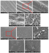
Representative scanning electron microscopy (SEM) micrographs of PTFE membranes: (a,b) PTFE-B (polytetrafluoroethylene-blue) side 1, showing areas with and without macro-waviness, and fine fibers distributed over them; (ce) PTFE-B side 2, shown with different magnification; crystallites in a form of dendrites are visible as a dominant nano-structural feature; fine fibers are not visible on this side. (fh) PTFE-W (polytetrafluoroethylene-white) surfaces, revealing heterogeneously distributed spherical crystallites or “warts” on an otherwise smooth surface; (i,j) PTFE-W micrographs showing the coexistence of “warts” (yellow arrow) and dendrites (red arrow).
Figure 5
Figure 5
Total surface free energy of bacteria and each side of the PTFE membranes calculated using the Owens–Wendt (O–W), Wu, and acid–base (A–B) model. PTFE-B (polytetrafluoroethylene-blue); PTFE-W (polytetrafluoroethylene-white).
Figure 6
Figure 6
(A) Bacterial adhesion to hexane, chloroform and diethyl ether, demonstrating their hydrophobic, electron-donor (Lewis base) and electron-acceptor (Lewis acid) character. (B) Bacterial colonization after 4 h on uncoated PTFE-W (polytetrafluoroethylene-white) and PTFE-B (polytetrafluoroethylene-blue) surface, and on a surface coated with artificial saliva. (a–l) Comparison within each bacterium species and different surfaces showed that the differences were statistically significant (p > 0.05).
Figure 7
Figure 7
Representative SEM micrographs of bacteria adhering to PTFE membranes. (ac) different magnifications of the corrugated area of PTFE-B (polytetrafluoroethylene-blue) membrane, containing thread-like structures; (d,e) bacteria adhering to PTFE-B in an area without thread-like structures; (fh) different magnifications of “warts”-rich area of PTFE-W (polytetrafluoroethylene-white) membrane; (i,j) bacteria adhering on areas without “warts”.

Similar articles

Cited by

References

    1. Morra M., Cassinelli C. Bacterial Adhesion to Polymer Surfaces: A Critical Review of Surface Thermodynamic Approaches. J. Biomater. Sci. Polym. Ed. 1998;9:55–74. doi: 10.1163/156856297X00263. - DOI - PubMed
    1. Teughels W., Van Assche N., Sliepen I., Quirynen M. Effect of Material Characteristics and/or Surface Topography on Biofilm Development. Clin. Oral Implants Res. 2006;17:68–81. doi: 10.1111/j.1600-0501.2006.01353.x. - DOI - PubMed
    1. Sterzenbach T., Helbig R., Hannig C., Hannig M. Bioadhesion in the Oral Cavity and Approaches for Biofilm Management by Surface Modifications. Clin. Oral Investig. 2020;24:4237–4260. doi: 10.1007/s00784-020-03646-1. - DOI - PMC - PubMed
    1. Lorenzetti M., Dogša I., Stošicki T., Stopar D., Kalin M., Kobe S., Novak S. The Influence of Surface Modification on Bacterial Adhesion to Titanium-Based Substrates. ACS Appl. Mater. Interfaces. 2015;7:1644–1651. doi: 10.1021/am507148n. - DOI - PubMed
    1. Zheng S., Bawazir M., Dhall A., Kim H.-E., He L., Heo J., Hwang G. Implication of Surface Properties, Bacterial Motility, and Hydrodynamic Conditions on Bacterial Surface Sensing and Their Initial Adhesion. Front. Bioeng. Biotechnol. 2021;9:643722. doi: 10.3389/fbioe.2021.643722. - DOI - PMC - PubMed

LinkOut - more resources