

Alteration of Oral and Perioral Soft Tissue in Mice following Incisor Tooth Extraction
- PMID: 35328407
- PMCID: PMC8951366
- DOI: 10.3390/ijms23062987
Alteration of Oral and Perioral Soft Tissue in Mice following Incisor Tooth Extraction
Abstract
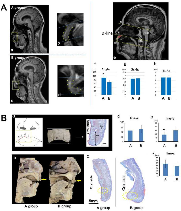
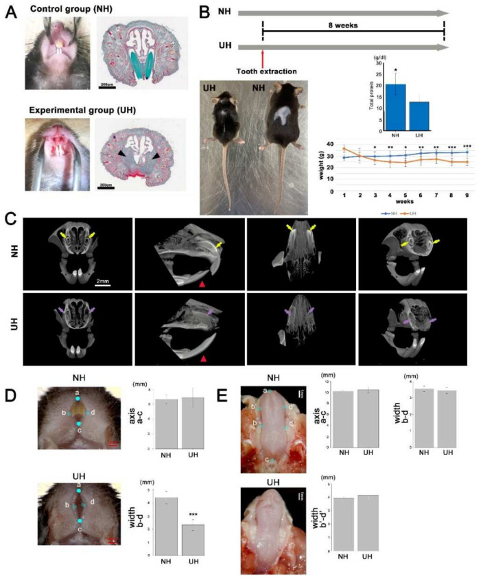
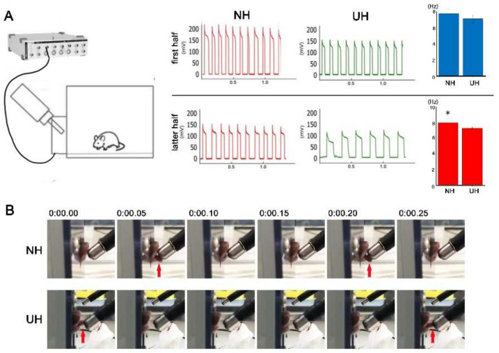
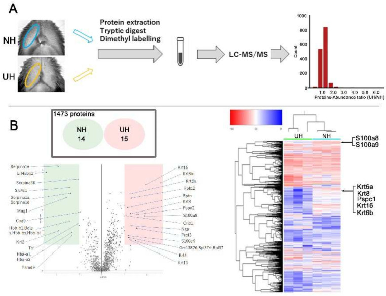
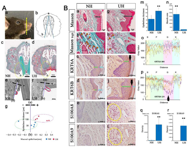
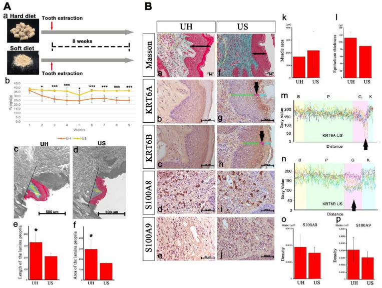
Oral and perioral soft tissues cooperate with other oral and pharyngeal organs to facilitate mastication and swallowing. It is essential for these tissues to maintain their morphology for efficient function. Recently, it was reported that the morphology of oral and perioral soft tissue can be altered by aging or orthodontic treatment. However, it remains unclear whether tooth loss can alter these tissues' morphology. This study examined whether tooth loss could alter lip morphology. First, an analysis of human anatomy suggested that tooth loss altered lip morphology. Next, a murine model of tooth loss was established by extracting an incisor; micro-computed tomography revealed that a new bone replaced the extraction socket. Body weight was significantly lower in the tooth loss (UH) group than in the non-extraction control (NH) group. The upper lip showed a greater degree of morphological variation in the UH group. Proteomic analysis and immunohistochemical staining of the upper lip illustrated that S100A8/9 expression was higher in the UH group, suggesting that tooth loss induced lip inflammation. Finally, soft-diet feeding improved lip deformity associated with tooth loss, but not inflammation. Therefore, soft-diet feeding is essential for preventing lip morphological changes after tooth loss.
Keywords: anatomy; human; lip morphology; mouse; tooth loss.
Conflict of interest statement
The authors declare no conflict of interest.
Figures
Similar articles
-
Measurement of three-dimensional perioral soft tissue changes in dentoalveolar protrusion patients after orthodontic treatment using a structured light scanner.Angle Orthod. 2014 Sep;84(5):795-802. doi: 10.2319/112913-877.1. Epub 2014 Mar 10. Angle Orthod. 2014. PMID: 24611593 Free PMC article.
-
Orthodontic incisor retraction caused changes in the soft tissue chin area: a retrospective study.BMC Oral Health. 2020 Apr 15;20(1):108. doi: 10.1186/s12903-020-01099-2. BMC Oral Health. 2020. PMID: 32295586 Free PMC article.
-
[Linear correlation between tooth movement and facial profile change in patients with class Ⅱ division 1 malocclusion].Zhonghua Kou Qiang Yi Xue Za Zhi. 2021 Jan 9;56(1):63-69. doi: 10.3760/cma.j.cn112144-20200330-00180. Zhonghua Kou Qiang Yi Xue Za Zhi. 2021. PMID: 34645237 Chinese.
-
Positional changes of the lips concomitant to incisor retraction: an important issue in orthodontics.World J Orthod. 2009 Fall;10(3):e9-11. World J Orthod. 2009. PMID: 21793260 Review.
-
Soft tissue changes following extraction vs. nonextraction orthodontic fixed appliance treatment: a systematic review and meta-analysis.Eur J Oral Sci. 2018 Jun;126(3):167-179. doi: 10.1111/eos.12409. Epub 2018 Feb 26. Eur J Oral Sci. 2018. PMID: 29480521
Cited by
-
Development and Regeneration of Muscle, Tendon, and Myotendinous Junctions in Striated Skeletal Muscle.Int J Mol Sci. 2022 Mar 10;23(6):3006. doi: 10.3390/ijms23063006. Int J Mol Sci. 2022. PMID: 35328426 Free PMC article. Review.
-
Effects of Myostatin on Nuclear Morphology at the Myotendinous Junction.Int J Mol Sci. 2023 Apr 2;24(7):6634. doi: 10.3390/ijms24076634. Int J Mol Sci. 2023. PMID: 37047606 Free PMC article.
-
Anatomical study of pterygoid implants: artery and nerve passage through bone dehiscence of the greater palatine canal.Int J Implant Dent. 2024 Nov 7;10(1):51. doi: 10.1186/s40729-024-00560-z. Int J Implant Dent. 2024. PMID: 39508991 Free PMC article.
References
-
- Desvarieux M., Demmer R.T., Rundek T., Boden-Albala B., Jacobs D.R., Papapanou P.N., Sacco R.L., Oral Infections and Vascular Disease Epidemiology Study (INVEST) Relationship between periodontal disease, tooth loss, and carotid artery plaque: The Oral Infections and Vascular Disease Epidemiology Study (INVEST) Stroke. 2003;34:2120–2125. doi: 10.1161/01.STR.0000085086.50957.22. - DOI - PMC - PubMed
MeSH terms
LinkOut - more resources
Full Text Sources
