. 2022 Mar 10;23(6):2994.
doi: 10.3390/ijms23062994.

Compartmentalized Innate Immune Response of Human Fetal Membranes against Escherichia coli Choriodecidual Infection

Affiliations

Compartmentalized Innate Immune Response of Human Fetal Membranes against Escherichia coli Choriodecidual Infection

Andrea Olmos-Ortiz et al. Int J Mol Sci. .

Abstract

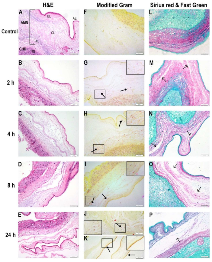
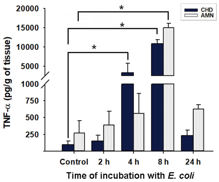
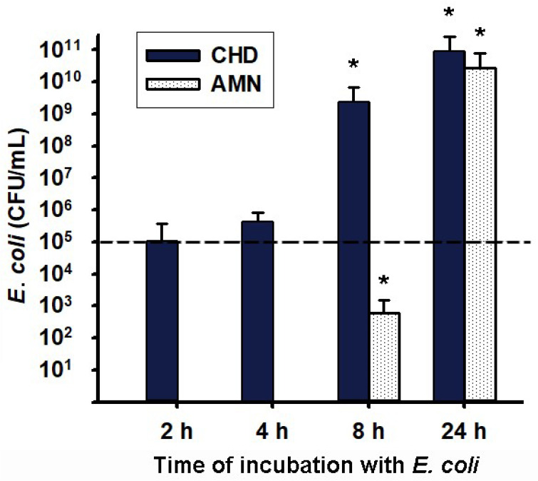
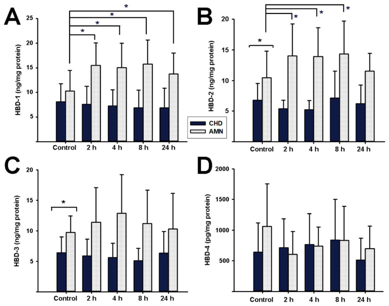
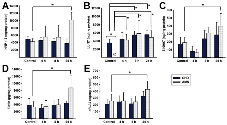
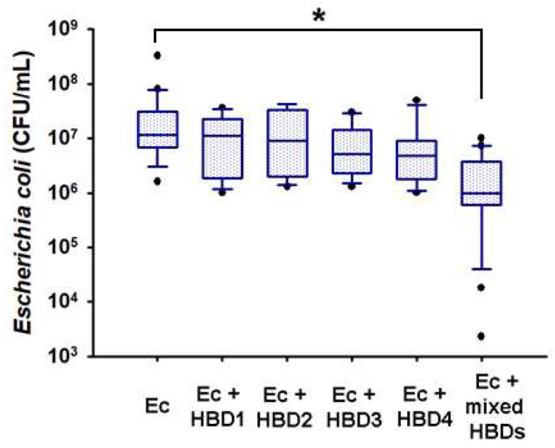
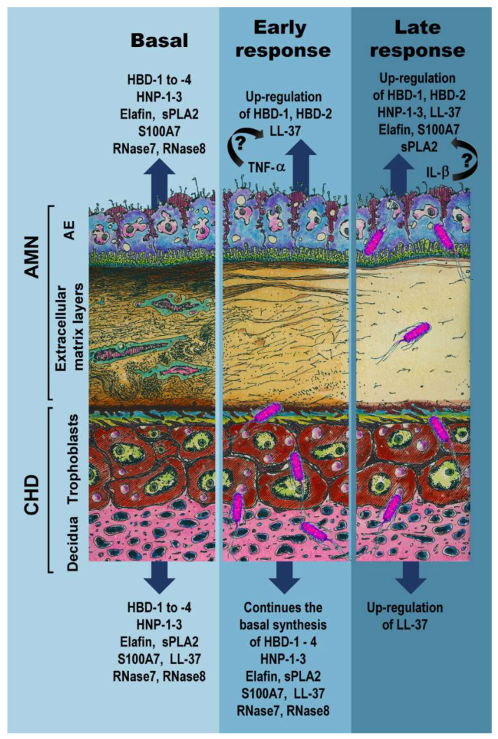
An infectious process into the uterine cavity represents a major endangered condition that compromises the immune privilege of the maternal-fetal unit and increases the risk for preterm birth (PTB) and premature rupture of membranes (PROM). Fetal membranes are active secretors of antimicrobial peptides (AMP), which limit bacterial growth, such as Escherichia coli. Nevertheless, the antibacterial responses displayed by chorioamniotic membranes against a choriodecidual E. coli infection have been briefly studied. The objective of this research was to characterize the profile of synthesis, activity, and spatial distribution of a broad panel of AMPs produced by fetal membranes in response to E. coli choriodecidual infection. Term human chorioamniotic membranes were mounted in a two independent compartment model in which the choriodecidual region was infected with live E. coli (1 × 10⁵ CFU/mL). Amnion and choriodecidual AMP tissue levels and TNF-α and IL-1β secretion were measured by the enzyme-linked immunosorbent assay. The passage of bacterium through fetal membranes and their effect on structural continuity was followed for 24 h. Our results showed that E. coli infection caused a progressive mechanical disruption of the chorioamniotic membranes and an activated inflammatory environment. After the challenge, the amnion quickly (2-4 h) induced production of human beta defensins (HBD)-1, HBD-2, and LL-37. Afterwards (8-24 h), the amnion significantly produced HBD-1, HBD-2, HNP-1-3, S100A7, sPLA2, and elafin, whereas the choriodecidua induced LL-37 synthesis. Therefore, we noticed a temporal- and tissue-specific pattern regulation of the synthesis of AMPs by infected fetal membranes. However, fetal membranes were not able to contain the collagen degradation or the bacterial growth and migration despite the battery of produced AMPs, which deeply increases the risk for PTB and PROM. The mixture of recombinant HBDs at low concentrations resulted in increased bactericidal activity compared to each HBD alone in vitro, encouraging further research to study AMP combinations that may offer synergy to control drug-resistant infections in the perinatal period.

Keywords: alpha defensins; antimicrobial peptides; beta defensins; collagen degradation; genitourinary infection; innate defense; innate immunity; maternal–fetal interface.


Conflict of interest statement

The authors declare that there is no conflict of interest regarding the publication of this paper.

Figures

Figure 1
Figure 1
Kinetic time follow-up of choriodecidual infection by Escherichia coli. The histological examination of E. coli-infected membranes was analyzed 2, 4, 8, and 24 h after tissue inoculation to evaluate the mechanical damage and bacteria migratory capacity through the CHD and AMN layers. Left panel (AE): Fetal membranes were stained with hematoxylin and eosin. Middle panel (FK): Modified Gram staining. The closed arrowheads show bacterial accumulation. Panel amplification is shown in the square. Right panel (LP): Collagen fibers were stained with Sirius red to observe zones with collagen degradation. The open arrowheads show zones with extracellular matrix degradation. Scale bar at 100 μm is shown at bottom right in each picture. Representative photographs from five independent experiments are shown. AE: amniotic epithelium. AMN: amnion. BL: Basal layer. CHD: choriodecidua. CL: Compact layer. FL: Fibroblastic layer. TB: Trophoblasts.
Figure 2
Figure 2
Pattern of TNF-α secretion into the culture medium from the chorioamniotic membranes infected with Escherichia coli. An inoculum of 1 × 10⁵ CFU/mL E. coli was used to infect the CHD compartment. Tissue culture proceeded for 24 h, and an aliquot of the medium was used to assess the TNF-α concentration at each follow-up time point (control, 2, 4, 8, and 24 h). The data are presented as means ± SD. n = 3 independent experiments in duplicate. * p < 0.05. AMN: Amnion. CHD: Choriodecidua.
Figure 3
Figure 3
Transmembrane migration of Escherichia coli from choriodecidual to amnion culture media. An inoculum of 1 × 10⁵ CFU/mL E. coli was used to infect the CHD compartment (shown as a dashed line). The tissue culture proceeded for 24 h, and an aliquot of the media was used to assess the CFU/mL at each follow-up time point (2, 4, 8, and 24 h). The data are presented as means ± SD. n = 6–7 independent experiments in triplicate. In the case of the CHD (dark bars), * p < 0.05 vs. the initial load of bacteria, represented by the dashed line. In the case of the AMN (white bars), * p < 0.05 vs. 2 h and 4 h, which were time points with undetectable bacteria, given that the inoculum was placed in the CHD compartment. As depicted, the bacteria migrated towards the AMN and were significantly detected at 8 h and 24 h. AMN: Amnion. CHD: Choriodecidua.
Figure 4
Figure 4
Effect of choriodecidual infection with Escherichia coli upon human beta defensins synthesis. An inoculum of 1 × 10⁵ CFU/mL E. coli was used to infect the CHD compartment. The tissue layers were lysed to assess HBD content at each follow-up time point (control, 2, 4, 8, and 24 h). (A) HBD-1, (B) HBD-2, (C) HBD-3, and (D) HBD-4. Beta defensin synthesis by the amnion (AMN) is shown in white bars, whereas synthesis by the choriodecidua (CHD) is shown in dark bars. The data are presented as means ± SD. n = 6 independent experiments in duplicate. * p < 0.05.
Figure 5
Figure 5
Effect of choriodecidual infection with Escherichia coli upon antimicrobial peptide synthesis. An inoculum of 1 × 10⁵ CFU/mL E. coli was used to infect the CHD compartment. The tissue layers were lysed to evaluate the specified AMP content at each follow-up time point (control, 4, 8, and 24 h). (A) HNP-1-3, (B) LL-37, (C) S100A7, (D) Elafin, and (E) sPLA2. AMP synthesis by the amnion (AMN) is shown in white bars, whereas synthesis by the choriodecidua (CHD) is shown in blue bars. The data are presented as means ± SD. n = 4–5 independent experiments in duplicate. * p < 0.05. ND = non-detectable.
Figure 6
Figure 6
Microbicidal effect of recombinant HBDs upon Escherichia coli growth. A constant load of bacteria (1 × 10⁵ CFU/mL) was incubated in the presence of 2000 ng/mL of each recombinant defensin alone or combined (500 ng/mL HBD-1 + 500 ng/mL HBD-2 + 500 ng/mL HBD-3 + 500 ng/mL HBD-4) in antibiotic-free medium. Incubations proceeded at 37 °C for 4 h. The E. coli control represents the normal bacteria growth in the absence of antimicrobial peptides. The data are presented as boxes (25th, 50th, and 75th percentile) and whiskers (5th and 95th percentile). n = 4–8 independent experiments in triplicate. Ec = Escherichia coli. * p < 0.05.
Figure 7
Figure 7
Schematic overview of AMP synthesis and the mechanical structure of chorioamniotic membranes in response to an Escherichia coli choriodecidual infection. In the basal state, the chorioamniotic membrane presents fully intact fibers of collagen. The AMN and CHD basally synthesize HBD-1 to -4, HNP-1-3, elafin, sPLA2, S100A7, RNAse-7, and RNAse-8, whereas LL-37 is basally absent in AMN. The challenge with a choriodecidual infection by E. coli disrupts the mechanical integrity of the collagen fibers and favors a TNF-α inflammatory milieu. The AMN side induces the early (2–4 h) synthesis of HBD-1, HBD-2, and LL-37 in response to E. coli. We hypothesize that TNF-α may drive the first AMP wave. At this time point, the CHD continues synthesizing all basally produced peptides. Bacteria bodies are observed only in the CHD compartment. In the late response (after 8 h of infection with E. coli), the extracellular matrix of the AMN was practically destroyed, and it appears to be an inflammatory environment enriched by IL-1β instead TNF-α. In this late wave after bacterial challenge, AMN synthesis of HBD-1, HBD-2, HNP-1-3, LL-37, S100A7, elafin, and sPLA2 are induced, whereas LL-37 is upregulated in the CHD compartment. IL-1β may control this observed second AMP wave. By this time, bacteria reach the amniotic compartment. AE: amniotic epithelium. AMN: amnion. CHD: choriodecidua.

Similar articles

Cited by

References

    1. Chawanpaiboon S., Vogel J.P., Moller A.-B., Lumbiganon P., Petzold M., Hogan D., Landoulsi S., Jampathong N., Kongwattanakul K., Laopaiboon M., et al. Global, Regional, and National Estimates of Levels of Preterm Birth in 2014: A Systematic Review and Modelling Analysis. Lancet Glob. Health. 2019;7:e37–e46. doi: 10.1016/S2214-109X(18)30451-0. - DOI - PMC - PubMed
    1. Crump C. Preterm Birth and Mortality in Adulthood: A Systematic Review. J. Perinatol. 2020;40:833–843. doi: 10.1038/s41372-019-0563-y. - DOI - PMC - PubMed
    1. McDonald H.M., Chambers H.M. Intrauterine Infection and Spontaneous Midgestation Abortion: Is the Spectrum of Microorganisms Similar to That in Preterm Labor? Infect. Dis. Obstet. Gynecol. 2000;8:220–227. doi: 10.1002/1098-0997(2000)8:5<220::AID-IDOG1022>3.0.CO;2-I. - DOI - PMC - PubMed
    1. Sáez-López E., Guiral E., Fernández-Orth D., Villanueva S., Goncé A., López M., Teixidó I., Pericot A., Figueras F., Palacio M., et al. Vaginal versus Obstetric Infection Escherichia coli Isolates among Pregnant Women: Antimicrobial Resistance and Genetic Virulence Profile. PLoS ONE. 2016;11:e0146531. doi: 10.1371/journal.pone.0146531. - DOI - PMC - PubMed
    1. Dautt-Leyva J.G., Canizalez-Román A., Acosta-Alfaro L.F., Gonzalez-Ibarra F., Murillo-Llanes J. Maternal and Perinatal Complications in Pregnant Women with Urinary Tract Infection Caused by Escherichia coli. J. Obstet. Gynaecol. Res. 2018;44:1384–1390. doi: 10.1111/jog.13687. - DOI - PubMed