

Emerging Role of HDACs in Regeneration and Ageing in the Peripheral Nervous System: Repair Schwann Cells as Pivotal Targets
- PMID: 35328416
- PMCID: PMC8951080
- DOI: 10.3390/ijms23062996
Emerging Role of HDACs in Regeneration and Ageing in the Peripheral Nervous System: Repair Schwann Cells as Pivotal Targets
Abstract
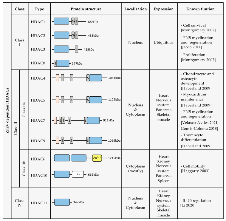
The peripheral nervous system (PNS) has a remarkable regenerative capacity in comparison to the central nervous system (CNS), a phenomenon that is impaired during ageing. The ability of PNS axons to regenerate after injury is due to Schwann cells (SC) being reprogrammed into a repair phenotype called Repair Schwann cells. These repair SCs are crucial for supporting axonal growth after injury, myelin degradation in a process known as myelinophagy, neurotropic factor secretion, and axonal growth guidance through the formation of Büngner bands. After regeneration, repair SCs can remyelinate newly regenerated axons and support nonmyelinated axons. Increasing evidence points to an epigenetic component in the regulation of repair SC gene expression changes, which is necessary for SC reprogramming and regeneration. One of these epigenetic regulations is histone acetylation by histone acetyl transferases (HATs) or histone deacetylation by histone deacetylases (HDACs). In this review, we have focused particularly on three HDAC classes (I, II, and IV) that are Zn2+-dependent deacetylases. These HDACs are important in repair SC biology and remyelination after PNS injury. Another key aspect explored in this review is HDAC genetic compensation in SCs and novel HDAC inhibitors that are being studied to improve nerve regeneration.
Keywords: HDACs; HDACs therapies; Schwann cell; ageing; myelin; nerve injury; nerve regeneration; remyelination; repair Schwann cell.
Conflict of interest statement
The authors declare no conflict of interest.
Figures
Similar articles
-
Sodium phenylbutyrate inhibits Schwann cell inflammation via HDAC and NFκB to promote axonal regeneration and remyelination.J Neuroinflammation. 2021 Oct 16;18(1):238. doi: 10.1186/s12974-021-02273-1. J Neuroinflammation. 2021. PMID: 34656124 Free PMC article.
-
Histone Acetylation in Central and Peripheral Nervous System Injuries and Regeneration: Epigenetic Dynamics and Therapeutic Perspectives.Int J Mol Sci. 2025 Jun 29;26(13):6277. doi: 10.3390/ijms26136277. Int J Mol Sci. 2025. PMID: 40650056 Free PMC article. Review.
-
Functions of histone modifications and histone modifiers in Schwann cells.Glia. 2020 Aug;68(8):1584-1595. doi: 10.1002/glia.23795. Epub 2020 Feb 8. Glia. 2020. PMID: 32034929 Review.
-
A Subpopulation of Foxj1-Expressing, Nonmyelinating Schwann Cells of the Peripheral Nervous System Contribute to Schwann Cell Remyelination in the Central Nervous System.J Neurosci. 2018 Oct 24;38(43):9228-9239. doi: 10.1523/JNEUROSCI.0585-18.2018. Epub 2018 Sep 18. J Neurosci. 2018. PMID: 30228229 Free PMC article.
-
After Nerve Injury, Lineage Tracing Shows That Myelin and Remak Schwann Cells Elongate Extensively and Branch to Form Repair Schwann Cells, Which Shorten Radically on Remyelination.J Neurosci. 2017 Sep 13;37(37):9086-9099. doi: 10.1523/JNEUROSCI.1453-17.2017. Epub 2017 Aug 3. J Neurosci. 2017. PMID: 28904214 Free PMC article.
Cited by
-
Isoviolanthin promotes Schwann cells activity in peripheral nerve regeneration via Fhl3-mediated epithelial-mesenchymal transition-like process: An in vitro study.Heliyon. 2024 Dec 12;11(1):e41087. doi: 10.1016/j.heliyon.2024.e41087. eCollection 2025 Jan 15. Heliyon. 2024. PMID: 39811297 Free PMC article.
-
Mesenchymal stem cell-based therapy for peripheral nerve injuries: A promise or reality?World J Stem Cells. 2025 Jun 26;17(6):107833. doi: 10.4252/wjsc.v17.i6.107833. World J Stem Cells. 2025. PMID: 40585952 Free PMC article. Review.
-
Histone Deacetylase 6 Inhibitor 5-Phenylcarbamoylpentyl Selenocyanide (SelSA) Suppresses Hepatocellular Carcinoma by Downregulating Phosphorylation of the Extracellular Signal-Regulated Kinase 1/2 Pathway.ACS Pharmacol Transl Sci. 2024 Jun 18;7(7):2196-2203. doi: 10.1021/acsptsci.4c00255. eCollection 2024 Jul 12. ACS Pharmacol Transl Sci. 2024. PMID: 39022367 Free PMC article.
-
rTMS ameliorates cerebral ischemia-reperfusion injury by inhibiting Golgi apparatus stress through epigenetic modulation of Gli2.Commun Biol. 2025 Aug 13;8(1):1209. doi: 10.1038/s42003-025-08613-8. Commun Biol. 2025. PMID: 40804340 Free PMC article.
-
Neuron-Schwann cell interactions in peripheral nervous system homeostasis, disease, and preclinical treatment.Front Cell Neurosci. 2023 Oct 12;17:1248922. doi: 10.3389/fncel.2023.1248922. eCollection 2023. Front Cell Neurosci. 2023. PMID: 37900588 Free PMC article. Review.
References
-
- Arthur-Farraj P.J., Latouche M., Wilton D.K., Quintes S., Chabrol E., Banerjee A., Woodhoo A., Jenkins B., Rahman M., Turmaine M., et al. c-Jun reprograms Schwann cells of injured nerves to generate a repair cell essential for regeneration. Neuron. 2012;75:633–647. doi: 10.1016/j.neuron.2012.06.021. - DOI - PMC - PubMed
-
- Jessen K.R., Mirsky R. Peripheral Nerve Tissue Engineering and Regeneration. Springer International Publishing; Berlin/Heidelberg, Germany: 2020. Schwann Cells in Nerve Repair and Regeneration; pp. 1–17.
Publication types
MeSH terms
Substances
Grants and funding
LinkOut - more resources
Full Text Sources
