. 2022 Mar 10;23(6):3005.
doi: 10.3390/ijms23063005.

The miRNA-21-5p Payload in Exosomes from M2 Macrophages Drives Tumor Cell Aggression via PTEN/Akt Signaling in Renal Cell Carcinoma

Affiliations

The miRNA-21-5p Payload in Exosomes from M2 Macrophages Drives Tumor Cell Aggression via PTEN/Akt Signaling in Renal Cell Carcinoma

Zhicheng Zhang et al. Int J Mol Sci. .

Abstract

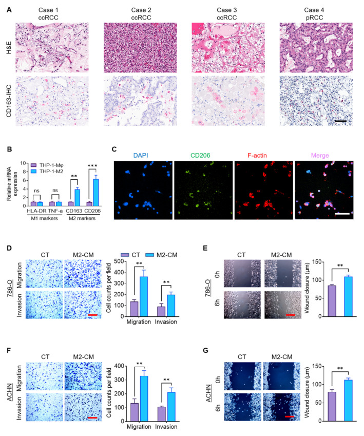
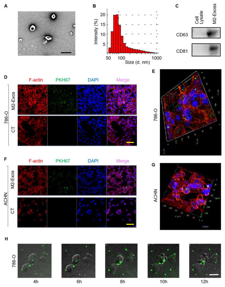
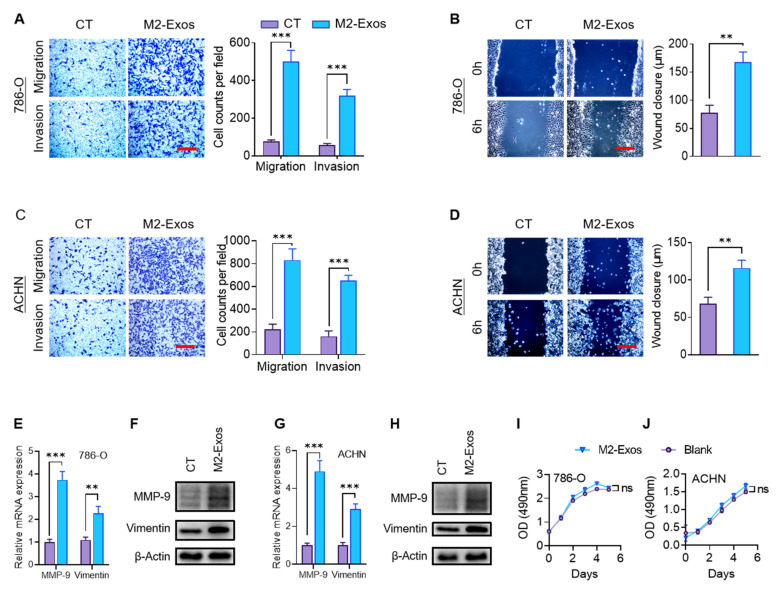
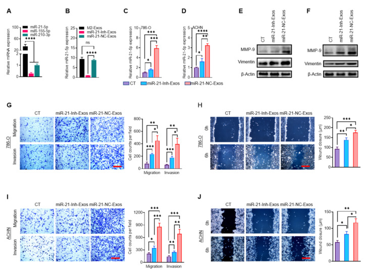
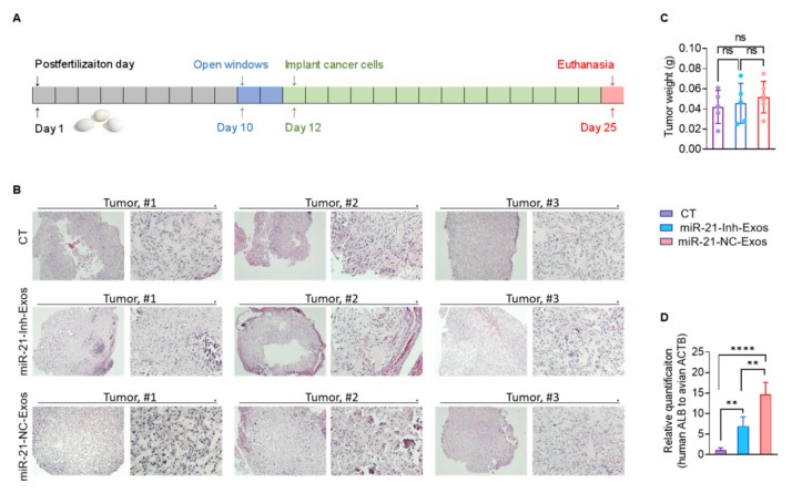
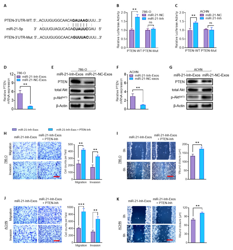
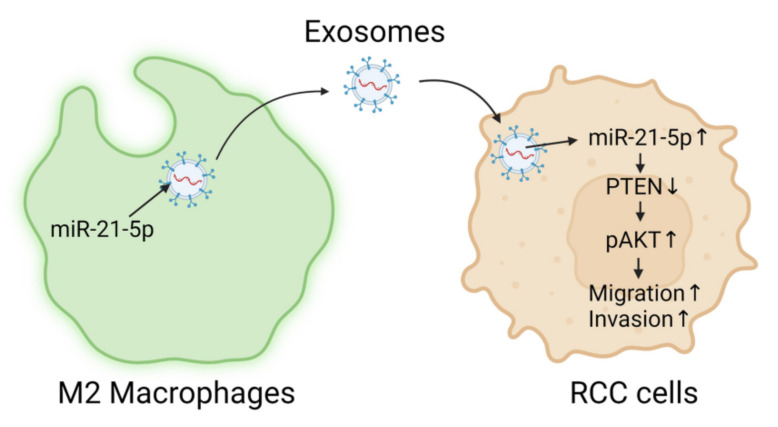
M2 macrophages in the tumor microenvironment are important drivers of cancer metastasis. Exosomes play a critical role in the crosstalk between different cells by delivering microRNAs or other cargos. Whether exosomes derived from pro-tumorigenic M2 macrophages (M2-Exos) could modulate the metastatic behavior of renal cell carcinoma (RCC) is unclear. This study found that M2-Exos promotes migration and invasion in RCC cells. Inhibiting miR-21-5p in M2-Exos significantly reversed their pro-metastatic effects on RCC cells in vitro and in the avian embryo chorioallantoic membrane in vivo tumor model. We further found that the pro-metastatic mechanism of miR-21-5p in M2-Exos is by targeting PTEN-3'UTR to regulate PTEN/Akt signaling. Taken together, our results demonstrate that M2-Exos carries miR-21-5p promote metastatic features of RCC cells through PTEN/Akt signaling. Reversing this could serve as a novel approach to control RCC metastasis.

Keywords: M2 macrophages; PTEN/Akt; exosomes; invasion; miRNA-21-5p; migration; renal cell carcinoma.


Conflict of interest statement

The authors declare no conflict of interest.

Figures

Figure 1
Figure 1
M2 macrophages promote RCC migration and invasion. (A) H&E and CD163 IHC staining (red) of four representative clinical RCC samples. Scale bar: 50 μm. (B) Macrophage marker mRNA expression was assayed in THP-1-Mφ and THP-1-M2 cells by RT-qPCR. (C) IF staining of nuclei (DAPI), CD206, and F-actin in THP-1-M2 cells. Scale bar: 50 μm. (DG) Cellular migration and invasion of 786-O and ACHN cells treated with M2-CM or control media were assessed by transwell and wound-healing assays. Scale bar: 100 μm. ** p < 0.01, *** p < 0.001, ns: not significant. pRCC: papillary renal cell carcinoma; CT: control; RT-qPCR: reverse transcription-quantitative PCR.
Figure 2
Figure 2
Characterization and internalization of M2-Exos. (A) TEM image of M2-Exos. Scale bar: 200 nm. (B) DLS measurement of M2-Exos size. (C) Western blotting assay of exosomal markers in THP-1-M2 cellular lysate and M2-Exos preparation. (D,F) Fluorescence images of 786-O and ACHN cells treated with or without PKH67-labeled M2-Exos (green). Scale bar: 50 μm. (E,G) Three-dimensional confocal reconstruction of 786-O and ACHN cells treated with PKH67-labeled M2-Exos (green). (H) Fluorescence staining analyzing the internalization of M2-Exos by 786-O cells over 12 h. Scale bar: 10 μm. CT: control.
Figure 3
Figure 3
M2-Exos promote RCC migration and invasion. 786-O and ACHN cells treated with or without M2-Exos. (AD) The impact of the M2-Exos treatment on the migration and invasion capabilities of RCC cells was analyzed by transwell (A,C) and wound-healing assays (B,D). Scale bar: 100 μm. (E,F) The impact of M2-Exos treatment on RCC cell: mRNA (E,G) and protein expression (F,H) of MMP-9 and vimentin were assayed by RT-qPCR and western blotting, respectively. (I,J) The impact of M2-Exos treatment on cell proliferation was analyzed by MTT assay. ** p < 0.01, *** p < 0.001. CT: control.
Figure 4
Figure 4
MiR-21-5p in M2-Exos promotes RCC migration and invasion in vitro. (A) The expression of three miRNAs in THP-1-M2 cells was analyzed by RT-qPCR. (B) The level of miR-21-5p expression in M2-Exos, M2-miR-21-Inh-Exos, and M2-miR-21-NC-Exos was assessed by RT-qPCR. (C,D) The intracellular content of miR-21-5p in 786-O and ACHN cells treated with control media, miR-21-Inh-Exos, or miR-21-NC-Exos was assessed by RT-qPCR. (E,F) The level of MMP-9 and vimentin in treated cells was assessed by western blotting. (GJ) The migration and invasion of treated cells were assessed by transwell (G,I) and wound-healing assays (H,J). Scale bar: 100 μm. * p < 0.05, ** p < 0.01, *** p < 0.001, **** p < 0.0001, ns: not significant.
Figure 5
Figure 5
MiR-21-5p in M2-Exos directs RCC aggressive behaviors in vivo. (A) Schematic diagram of the timeline of CAM xenograft implantation. Three groups of 786-O tumor cells were implanted on CAMs following treatment with exosomes-free media (the control group), miR-21-Inh-Exos, or miR-21-NC-Exos. (B) H&E stains of three representative CAM-derived tumors from each group are shown. Magnification: 40×, 200×, for each tumor sample. (C) The tumor weights of harvested CAM tumors from each group. (D) qPCR for human ALB in embryonic livers normalized to avian ACTB. ** p < 0.01, **** p < 0.0001, ns: not significant.
Figure 6
Figure 6
MiR-21-5p in M2-Exos regulates PTEN/AKT signaling to promote RCC metastasis. (A) The sequences of the putative target site for miR-21-5p in the PTEN-3′UTR (PTEN-3′UTR-WT) and a mutated version of this target (PTEN-3′UTR-Mut) were shown. (B,C) 786-O and ACHN cells were co-transfected psiCHECK2-PTEN-3′UTR-WT or psiCHECK2-PTEN-3′UTR-Mut, with miR-21-Inh or miR-21-NC. The relative luciferase activities in the cells were assayed. (DG) The expression of PTEN mRNA, and protein level of PTEN, total Akt, and p-AktS473 were assessed in 786-O and ACHN cells treated with miR-21-Inh-Exos or miR-21-NC-Exos. (HK) The migration and invasive capabilities of cells treated with miR-21-Inh-Exos or miR-21-Inh-Exos + PTEN inhibitor (PTEN-Inh) were analyzed by transwell and wound-healing assays. Scale bar, 100 μm. ** p < 0.01, *** p < 0.001, ns: not significant.
Figure 7
Figure 7
Schematic diagram of the proposed mechanism of M2-Exos mediated enhancement of RCC aggression via miR-21-5p.

Similar articles

Cited by

References

    1. Capitanio U., Bensalah K., Bex A., Boorjian S.A., Bray F., Coleman J., Gore J.L., Sun M., Wood C., Russo P. Epidemiology of renal cell carcinoma. Eur. Urol. 2019;75:74–84. doi: 10.1016/j.eururo.2018.08.036. - DOI - PMC - PubMed
    1. Hsieh J.J., Purdue M.P., Signoretti S., Swanton C., Albiges L., Schmidinger M., Heng D.Y., Larkin J., Ficarra V. Renal cell carcinoma. Nat. Rev. Dis. Primers. 2017;3:17009. doi: 10.1038/nrdp.2017.9. - DOI - PMC - PubMed
    1. Tannir N.M., Pal S.K., Atkins M.B. Second-line treatment landscape for renal cell carcinoma: A comprehensive review. Oncologist. 2018;23:540–555. doi: 10.1634/theoncologist.2017-0534. - DOI - PMC - PubMed
    1. Pontes O., Oliveira-Pinto S., Baltazar F., Costa M. Renal cell carcinoma therapy: Current and new drug candidates. Drug Discov. Today. 2022;27:304–314. doi: 10.1016/j.drudis.2021.07.009. - DOI - PubMed
    1. Martinez F.O., Helming L., Gordon S. Alternative activation of macrophages: An immunologic functional perspective. Annu. Rev. Immunol. 2009;27:451–483. doi: 10.1146/annurev.immunol.021908.132532. - DOI - PubMed

MeSH terms