. 2022 Mar 11;23(6):3024.
doi: 10.3390/ijms23063024.

Identifying Function Determining Residues in Neuroimmune Semaphorin 4A

Affiliations

Identifying Function Determining Residues in Neuroimmune Semaphorin 4A

Svetlana P Chapoval et al. Int J Mol Sci. .

Abstract

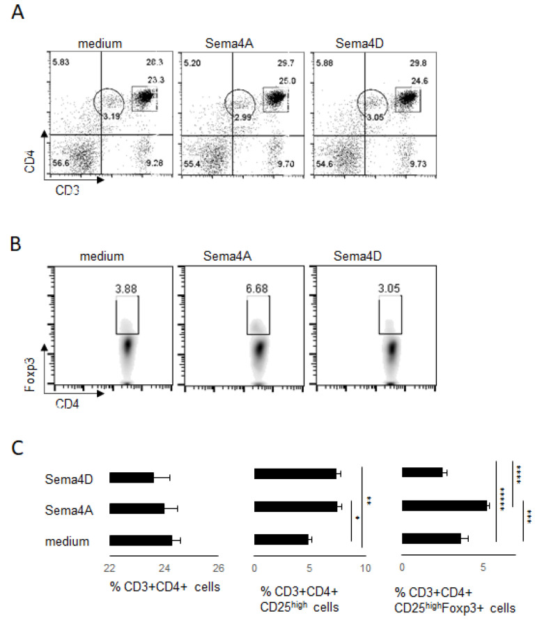
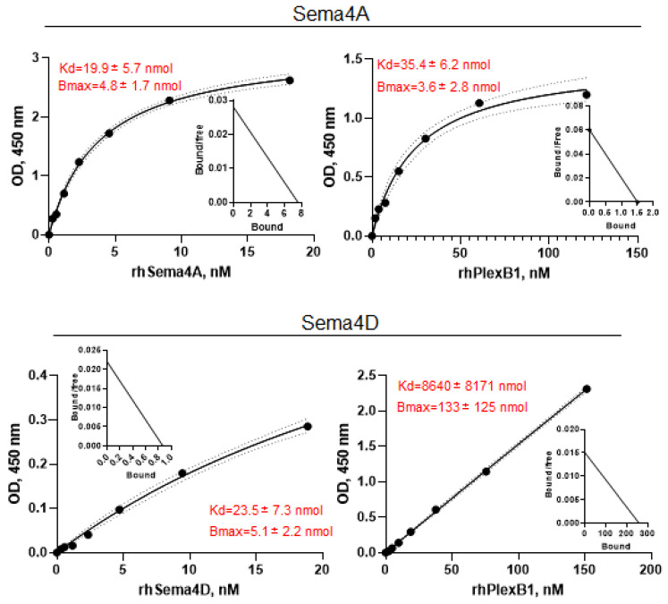
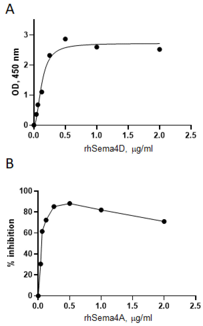
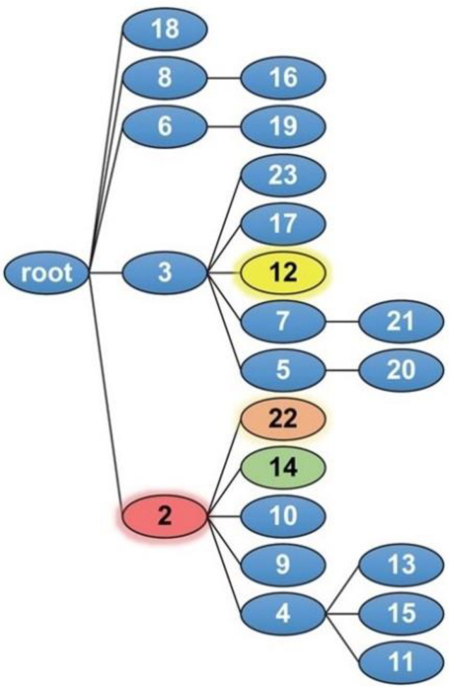
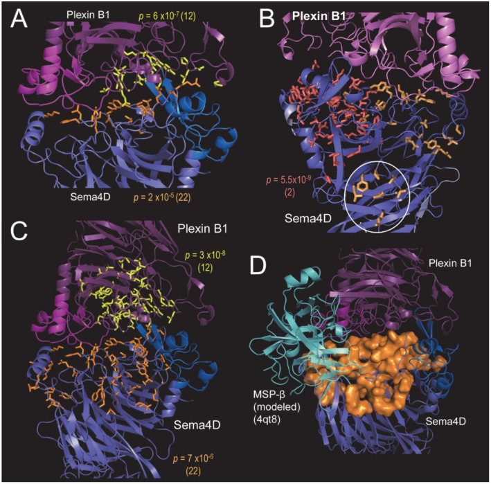
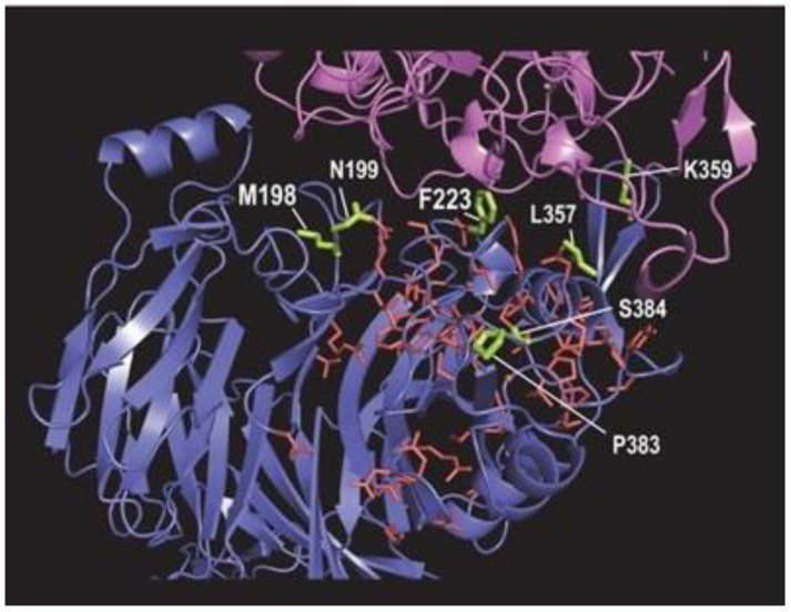
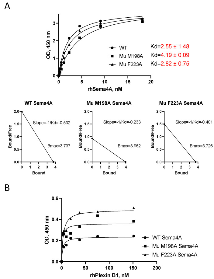
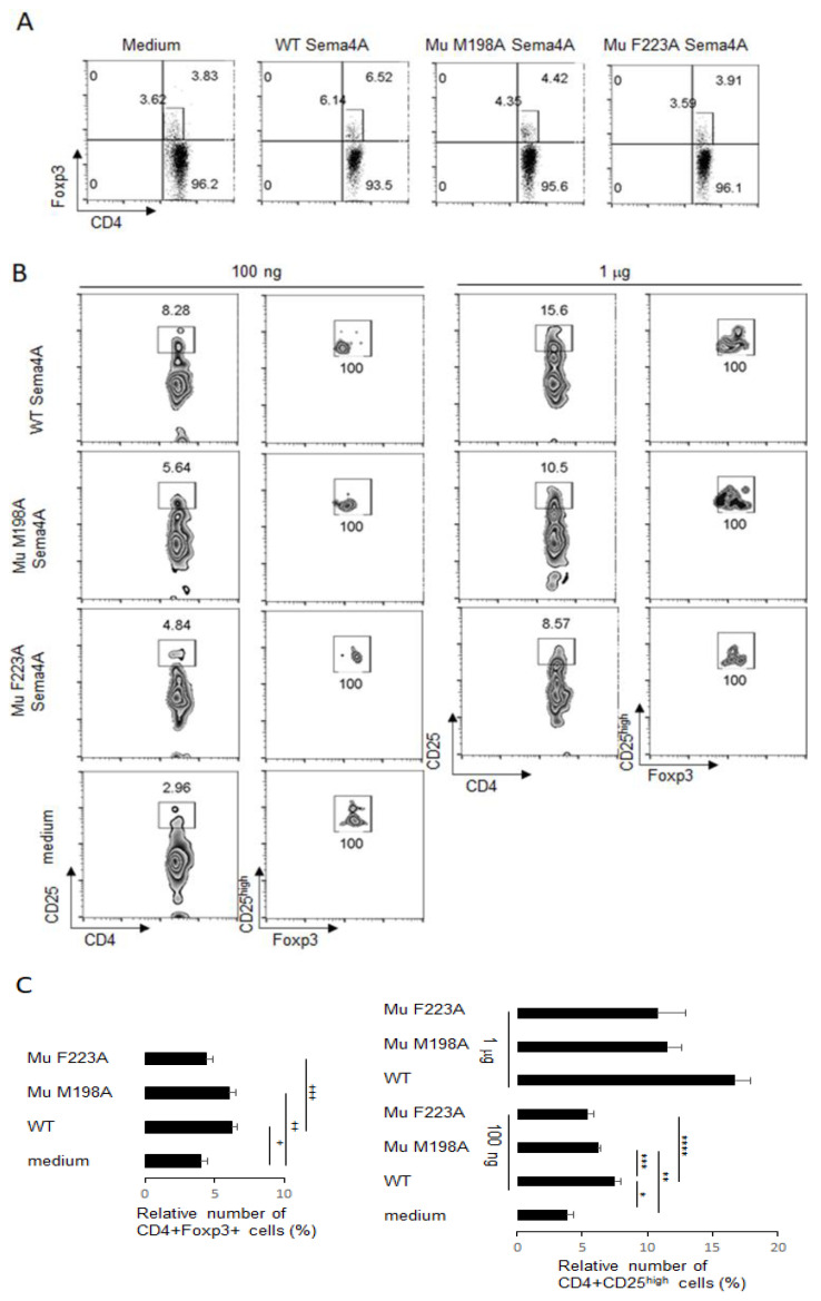
Semaphorin 4A (Sema4A) exerts a stabilizing effect on human Treg cells in PBMC and CD4+ T cell cultures by engaging Plexin B1. Sema4A deficient mice display enhanced allergic airway inflammation accompanied by fewer Treg cells, while Sema4D deficient mice displayed reduced inflammation and increased Treg cell numbers even though both Sema4 subfamily members engage Plexin B1. The main objectives of this study were: 1. To compare the in vitro effects of Sema4A and Sema4D proteins on human Treg cells; and 2. To identify function-determining residues in Sema4A critical for binding to Plexin B1 based on Sema4D homology modeling. We report here that Sema4A and Sema4D display opposite effects on human Treg cells in in vitro PBMC cultures; Sema4D inhibited the CD4+CD25+Foxp3+ cell numbers and CD25/Foxp3 expression. Sema4A and Sema4D competitively bind to Plexin B1 in vitro and hence may be doing so in vivo as well. Bayesian Partitioning with Pattern Selection (BPPS) partitioned 4505 Sema domains from diverse organisms into subgroups based on distinguishing sequence patterns that are likely responsible for functional differences. BPPS groups Sema3 and Sema4 into one family and further separates Sema4A and Sema4D into distinct subfamilies. Residues distinctive of the Sema3,4 family and of Sema4A (and by homology of Sema4D) tend to cluster around the Plexin B1 binding site. This suggests that the residues both common to and distinctive of Sema4A and Sema4D may mediate binding to Plexin B1, with subfamily residues mediating functional specificity. We mutated the Sema4A-specific residues M198 and F223 to alanine; notably, F223 in Sema4A corresponds to alanine in Sema4D. Mutant proteins were assayed for Plexin B1-binding and Treg stimulation activities. The F223A mutant was unable to stimulate Treg stability in in vitro PBMC cultures despite binding Plexin B1 with an affinity similar to the WT protein. This research is a first step in generating potent mutant Sema4A molecules with stimulatory function for Treg cells with a view to designing immunotherapeutics for asthma.

Keywords: Plexin B1; Semaphorin 4A; human Treg cells; immunotherapeutics for asthma; mutated proteins.


Conflict of interest statement

The authors declare no conflict of interest.

Figures

Figure 1
Figure 1
Human Sema4 proteins affect Treg cells in PBMC cultures. Human PBMC were cultured with or without 100 ng of external rhSema4A or rhSema4D for 48 h. After harvest, the numbers of CD4+ T cells (panels (A,C)) and Treg cells (panels (B,C)) were assessed by flow cytometry, using specific Abs to corresponding cell surface and intracellular molecules. CD3+CD4+ cells shown on upper panel dot plots (A) were further selected to evaluate the relative number of Treg cells (B) in each in vitro experimental setting. The Treg cells were gated on CD4+Foxp3+ density plots. (C) The graphical representation of data with corresponding statistics. * p < 0.013, cultures with medium vs. rhSema4A, ** p < 0.012, medium vs. rhSema4D, *** p = 0.012, medium vs. rhSema4A, **** p = 0.001, rhSema4A vs. rhSema4D, ***** p = 0.040, medium vs. rhSema4D. Data are representative from three independent experiments.
Figure 2
Figure 2
Sema4A binds Plexin B1 with higher affinity than Sema4D. Analysis of Sema4A-Plexin B1 and Sema4D–Plexin B1 interaction by the direct ligand–receptor binding ELISAs with respective Scatchard plots shown as inserts. Either Plexin B1 (8–10 μg/mL) or Sema4 (2 μg/mL) were immobilized on the Immulon 2 plates overnight and corresponding ligands or receptor were added in duplicates in varying doses, starting from 2 μg/mL for Sema4 and 10 μg/mL for Plexin B1. The amount of bound ligand was measured spectrophotometrically based on the HRP-labeled Abs to corresponding protein tags (Fc, GST, or His). Calculations of the equilibrium dissociation constants were performed utilizing GraphPrizm 8.0. software. For the KD value determination, the data was fitted to the ‘One site-specific binding with Hill slope’. Data are shown in mean ± SEM from three independent ELISA experiments. The curves demonstrate a direct 1:1 ligand–receptor binding and are representative of three independent assays.
Figure 3
Figure 3
Sema4A and Sema4D compete for Plexin B1 binding in the in vitro ELISA assay. The HIS-Select nickel-coated microplates were covered with 10 μg/mL of Plexin B1 with His tag in Tris Buffer overnight. Either rhSema4A-GST or -Fc or rhSema4D-Fc in duplicates were applied to the plates in varying doses starting from 2 μg/mL. (A) rhSema4D binding to Plexin B1 in the direct LRA ELISA. (B) Inhibition of 2 μg/mL Sema4D binding to Plexin B1 by increasing concentrations of Sema4A, starting at 2 μg/mL dose. The OD for Sema4A+Sema4D was compared to the corresponding OD for Sema4D alone in the wells covered with Plexin B1 protein.
Figure 4
Figure 4
BPPS defined Sema domain hierarchy. Sema4A, Sema4D, and PlexinB1 correspond to nodes 14, 22, and 12, respectively.
Figure 5
Figure 5
BPPS-SIPRIS analyses of the Sema4D–PlexinB1 complex (pdb_id: 3ol2). (A) BPPS-SIPRIS analysis identifying Sema4D- and PlexinB1-specific residues significantly clustered at the Sema4D–PlexinB1 interface; p-values and, in parentheses, the corresponding node IDs are indicated. Shown in orange are Sema4D residues contacting Plexin B1 and in yellow Plexin B1 residues contacting Sema4D. (B) Sema4D family- and subfamily-specific residue clusters identified by BPPS-SIPRIS (residue sidechains shown in red and orange, respectively). Two subfamily clusters were found: adjacent to the Plexin B1 interface and another at the Sema4D homodimeric interface (circled). (C) BPPS-SIPRIS analysis identifying Sema4D- and PlexinB1-specific residues significantly clustered within each subunit. (D) The Sema4D-specific residues in panel C (orange space-fill) are relative to a modeled interaction with MSP-β subunit. Bound MSP-β was modeled by structurally superimposing, over Plexin B1, the Ron Sema domain + MSP-β complex (pdb_id: 4qt8). Although merely hypothetical, this suggests that the larger Sema4D surface in panel C may facilitate binding of other cellular components.
Figure 6
Figure 6
Modeling of Sema4A subfamily-specific residues (lime-colored sidechains) based on homology to the Sema4D structure. The sidechains of node 2 family residues (within Sema4D) are shown in red. The backbone of Plexin B1 is shown in pink and of Sema4D in blue.
Figure 7
Figure 7
WT and Mutant (Mu) Sema4A proteins bind Plexin B1 with similar affinity. Direct (A) and reverse (B) LRA ELISAs for WT and Mu Sema4A protein binding to Plexin B1 were performed as described in Materials and Methods, analyzed in GraphPrizm 8.0 software, and corresponding Kd determination was performed as described for Figure 2. Kd are shown in mean ± SEM from three independent ELISA experiments, whereas slopes are from one representative experiment.
Figure 8
Figure 8
Mutant Sema4A molecules are less potent in the stabilization of human peripheral Treg cells in vitro as compared to WT recombinant Sema4A protein. Human PBMC were cultured with or without 100 ng of external WT Sema4A or Mu Sema4A for 48 h. After harvest, the numbers of Treg cells (panels (A,C)) were assessed by flow cytometry, using specific Abs to corresponding cell surface and intracellular molecules. The Treg cells were gated on CD4 + Foxp3+ dot plots. The relative number of CD3 + CD4 + CD25high cells (panels (B,C)) for two distinct concentrations of individual recombinant protein in each in vitro experimental setting. Data are representative from two independent experiments. + p < 0.012, medium vs. WT Sema4A; ++ p < 0.02, medium vs. Mu M198A Sema4A; +++ p < 0.038, WT vs. Mu F223A Sema4A; * p < 0.002, med vs. WT Sema4A, ** p < 0.054, med vs. Mu M198A, *** p < 0.041, WT vs. Mu F198A Sema4A, **** p < 0.016, WT vs. F223A Sema4A.

Similar articles

Cited by

References

    1. Pasterkamp R.J., Giger R.J. Semaphorin Function in Neural Plasticity and Disease. Curr. Opin. Neurobiol. 2009;19:263–274. doi: 10.1016/j.conb.2009.06.001. - DOI - PMC - PubMed
    1. Rozbesky D., Robinson R., Jain V., Renner M., Malinauskas T., Harlos K., Siebold C., Jones E.Y. Diversity of Oligomerization in Drosophila Semaphorins Suggests a Mechanism of Functional Fine-Tuning. Nat. Commun. 2019;10:1–12. doi: 10.1038/s41467-019-11683-y. - DOI - PMC - PubMed
    1. Iyer A.S., Chapoval S.P. Neuroimmune Semaphorin 4A in Cancer Angiogenesis and Inflammation: A Promoter or a Suppressor? Int. J. Mol. Sci. 2018;20:124. doi: 10.3390/ijms20010124. - DOI - PMC - PubMed
    1. Adi S.D., Eiza N., Bejar J., Shefer H., Toledano S., Kessler O., Neufeld G., Toubi E., Vadasz Z. Semaphorin 3A Is Effective in Reducing Both Inflammation and Angiogenesis in a Mouse Model of Bronchial Asthma. Front. Immunol. 2019;10:550. doi: 10.3389/fimmu.2019.00550. - DOI - PMC - PubMed
    1. Tatari N., Movassagh H., Shan L., Koussih L., Gounni A.S. Semaphorin 3E Inhibits House Dust Mite–Induced Angiogenesis in a Mouse Model of Allergic Asthma. Am. J. Pathol. 2019;189:762–772. doi: 10.1016/j.ajpath.2019.01.008. - DOI - PubMed

LinkOut - more resources