. 2022 Mar 11;23(6):3027.
doi: 10.3390/ijms23063027.

Influence of Urea and Dimethyl Sulfoxide on K-Peptide Fibrillation

Affiliations

Influence of Urea and Dimethyl Sulfoxide on K-Peptide Fibrillation

Jarosław Wawer et al. Int J Mol Sci. .

Abstract

Protein fibrillation leads to formation of amyloids-linear aggregates that are hallmarks of many serious diseases, including Alzheimer's and Parkinson's diseases. In this work, we investigate the fibrillation of a short peptide (K-peptide) from the amyloidogenic core of hen egg white lysozyme in the presence of dimethyl sulfoxide or urea. During the studies, a variety of spectroscopic methods were used: fluorescence spectroscopy and the Thioflavin T assay, circular dichroism, Fourier-transform infrared spectroscopy, optical density measurements, dynamic light scattering and intrinsic fluorescence. Additionally, the presence of amyloids was confirmed by atomic force microscopy. The obtained results show that the K-peptide is highly prone to form fibrillar aggregates. The measurements also confirm the weak impact of dimethyl sulfoxide on peptide fibrillation and distinct influence of urea. We believe that the K-peptide has higher amyloidogenic propensity than the whole protein, i.e., hen egg white lysozyme, most likely due to the lack of the first step of amyloidogenesis-partial unfolding of the native structure. Urea influences the second step of K-peptide amyloidogenesis, i.e., folding into amyloids.

Keywords: K-peptide; amyloids; dimethyl sulfoxide; fibrillation; hen egg white lysozyme; urea.


Conflict of interest statement

The authors declare no conflict of interest.

Figures

Figure 1
Figure 1
The ThT florescence intensity recorded at 485 nm for the samples of K-peptide incubated at 37 °C up to 14 days. The peptide was dissolved in: ✕ water; □ acetate buffer, pH = 4, without additives; ■ acetate buffer with dimethyl sulfoxide and ● acetate buffer with urea. Symbol ▲ refers to blank, control solutions without the K-peptide. Experimental uncertainty is presented as standard deviation.
Figure 2
Figure 2
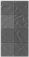
The atomic force microscopy scans for the samples of K-peptide in freshly prepared solutions (left column) and in the samples incubated at 37 °C for 14 days (right column). The peptide was dissolved in: (A,B) water; (C,D) acetate buffer, pH = 4, without additives; (E,F) acetate buffer with dimethyl sulfoxide and (G,H) acetate buffer with urea.
Figure 3
Figure 3
Size distribution by number obtained from dynamic light scattering measurements for the samples of K-peptide in freshly prepared solutions (dark blue line) and for the samples of K-peptide incubated at 37 °C for 14 days (red line) dissolved in: (A) water; (B) acetate buffer, pH = 4, without additives; (C) acetate buffer with dimethyl sulfoxide and (D) acetate buffer with urea.
Figure 4
Figure 4
Mean residue ellipticity (MRE) recorded for: (A) samples of non-incubated freshly prepared solutions of the K-peptide, (B) samples of the K-peptide incubated at 37 °C for 14 days. The peptide was dissolved in: water (blue line); acetate buffer, pH = 4, without additives (red line) and acetate buffer with urea (dark yellow line).
Figure 5
Figure 5
Infrared spectra—expressed as second derivatives of absorbance—recorded for: (A) samples of non-incubated freshly prepared solutions of the K-peptide and (B) samples of the K-peptide incubated at 37 °C for 14 days. The peptide was dissolved in: water (blue line); acetate buffer, pH = 4, without additives (red line) and acetate buffer with dimethyl sulfoxide (green line).
Figure 6
Figure 6
Intrinsic fluorescence of the K-peptide in freshly prepared solutions (dark blue line) and its fluorescence after incubation at 37 °C for 14 days (red line). The peptide was dissolved in: (A) water; (B) acetate buffer, pH = 4, without additives; (C) acetate buffer with dimethyl sulfoxide and (D) acetate buffer with urea.

Similar articles

References

    1. Chiti F., Dobson C.M. Protein Misfolding, Functional Amyloid, and Human Disease. Annu. Rev. Biochem. 2006;75:333–366. doi: 10.1146/annurev.biochem.75.101304.123901. - DOI - PubMed
    1. Makin O.S., Serpell L.C. Structures for amyloid fibrils. FEBS J. 2005;272:5950–5961. doi: 10.1111/j.1742-4658.2005.05025.x. - DOI - PubMed
    1. Hill S.E., Miti T., Richmond T., Muschol M. Spatial Extent of Charge Repulsion Regulates Assembly Pathways for Lysozyme Amyloid Fibrils. PLoS ONE. 2011;6:e18171. doi: 10.1371/journal.pone.0018171. - DOI - PMC - PubMed
    1. Sunde M., Serpell L.C., Bartlam M., Fraser P.E., Pepys M.B., Blake C.C. Common core structure of amyloid fibrils by synchrotron X-ray diffraction. J. Mol. Biol. 1997;273:729–739. doi: 10.1006/jmbi.1997.1348. - DOI - PubMed
    1. Arnaudov L.N., De Vries R. Thermally induced fibrillar aggregation of hen egg white lysozyme. Biophys. J. 2005;88:515–526. doi: 10.1529/biophysj.104.048819. - DOI - PMC - PubMed

LinkOut - more resources