. 2022 Mar 11;23(6):3032.
doi: 10.3390/ijms23063032.

Tuning Strain Stiffening of Protein Hydrogels by Charge Modification

Affiliations

Tuning Strain Stiffening of Protein Hydrogels by Charge Modification

Jie Gu et al. Int J Mol Sci. .

Abstract

Strain-stiffening properties derived from biological tissue have been widely observed in biological hydrogels and are essential in mimicking natural tissues. Although strain-stiffening has been studied in various protein-based hydrogels, effective approaches for tuning the strain-stiffening properties of protein hydrogels have rarely been explored. Here, we demonstrated a new method to tune the strain-stiffening amplitudes of protein hydrogels. By adjusting the surface charge of proteins inside the hydrogel using negatively/positively charged molecules, the strain-stiffening amplitudes could be quantitively regulated. The strain-stiffening of the protein hydrogels could even be enhanced 5-fold under high deformations, while the bulk property, recovery ability and biocompatibility remained almost unchanged. The tuning of strain-stiffening amplitudes using different molecules or in different protein hydrogels was further proved to be feasible. We anticipate that surface charge adjustment of proteins in hydrogels represents a general principle to tune the strain-stiffening property and can find wide applications in regulating the mechanical behaviors of protein-based hydrogels.

Keywords: electrical repulsion; mechanical property; protein hydrogel; strain-stiffening; surface charge.


Conflict of interest statement

The authors declare no conflict of interest.

Figures
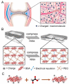
Figure 1
Figure 1
Illustration of tuning the strain-stiffening of BSA-PEG hydrogels by surface charge modification of proteins. (A) Schematic of cartilage with charged proteoglycans dispersed inside. (B) Compression–relaxation cycle of the BSA-PEG network and the electrical repulsion between adjacent BSA with charge modified under compression. (C) Surface charge modification of BSA using glyoxylic acid.
Figure 2
Figure 2
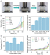
Compressive mechanical properties of BSA-PEG hydrogels. (A) Optical images of compression and relaxation of BSA-PEG hydrogels. (B) Typical stress–strain curves under compression for BSA-PEG hydrogels at different BSA:PEG ratios and solid contents in air. The concentrations of PEG and BSA were 6 and 5 mM, 8 and 5 mM, 10 and 5 mM, 10 and 8 mM, and 12 and 10 mM, respectively. (C,D) Young’s modulus (C) and toughness (D) correspond to BSA-PEG hydrogels at different BSA:PEG ratios and solid contents. (E) Differential modulus of BSA-PEG hydrogels at various strains.
Figure 3
Figure 3
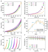
Surface charge modification of BSA and bulk properties of modified BSA-PEG hydrogels. (A,B) Zeta potential (A) and CD spectra (B) of modified BSA. Different molar ratios of glyoxylic acid and BSA were used in the modification (0:0, 1:1, 2:1, 2.5:1, 3:1 and 4:1, simplified as 0, 1.0, 2.0, 2.5, 3.0 and 4.0). (C) UV absorbance of DTNB-containing leachates of BSA-PEG hydrogels modified at different ratios of glyoxylic acid and BSA. The UV absorbance at 412 nm indicates the reaction products of DTNB, which were used to indicate the exposed thiol from unfolded BSA in hydrogels. (D) SEM images of BSA-PEG hydrogels modified at different ratios of glyoxylic acid and BSA.
Figure 4
Figure 4
Compressibility of the BSA-PEG hydrogels modified with glyoxylic acid. (A) Compressibility of BSA-PEG hydrogels modified at different ratios of glyoxylic acid and BSA. Different molar ratios of glyoxylic acid and BSA were used in the modification (0:0, 1:1, 2:1, 2.5:1, 3:1 and 4:1, simplified as 0, 1.0, 2.0, 2.5, 3.0 and 4.0). (B) Differential modulus corresponding to BSA-PEG hydrogels modified at different ratios of glyoxylic acid and BSA. (C) Compression–relaxation of modified BSA-PEG hydrogels at the strain of 50%. (D) Summarized stress for modified BSA-PEG hydrogels at the strain of 50% and zeta potentials of BSA modified at different ratios of glyoxylic acid and BSA. (E) Continuous compression–relaxation cycles of BSA-PEG hydrogels modified with different ratios of glyoxylic acid and BSA for 10 cycles. (F) Normalized maximum stress of BSA-PEG hydrogels modified at different ratios of glyoxylic acid and BSA in 10 cycles of compression and relaxation.
Figure 5
Figure 5
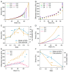
Mechanical properties of BSA-PEG hydrogels modified with bromoacetic acid and hemoglobin-PEG hydrogels modified with glyoxylic acid. (A) Compressibility of BSA-PEG hydrogels modified with bromoacetic acid. Different molar ratios of bromoacetic acid and BSA were used in the modification (0:0, 0.2:1, 0.4:1, 0.6:1, 0.8:1 and 1.0:1, simplified as 0, 0.2, 0.4, 0.6, 0.8 and 1.0). (B) Differential modulus corresponding to BSA-PEG hydrogels modified with different ratios of bromoacetic acid and BSA. (C) Summarized stress for modified BSA-PEG hydrogels at the strain of 50% and zeta potentials of BSA at different ratios of bromoacetic acid and BSA. (D) Compressibility of hemoglobin-PEG hydrogels modified with glyoxylic acid. Different molar ratios of glyoxylic acid: hemoglobin were used in the modification (0:0, 1:1 and 2:1, simplified as 0.0, 1.0 and 2.0). (E) Differential modulus corresponds to hemoglobin-PEG hydrogels modified at different ratios of glyoxylic acid and hemoglobin. (F) Summarized stress for modified hemoglobin-PEG hydrogels at the strain of 50% and zeta potentials of hemoglobin modified at different ratios of glyoxylic acid and hemoglobin.

Similar articles

Cited by

References

    1. Wang Y., Xu Z., Lovrak M., le Sage V.A.A., Zhang K., Guo X., Eelkema R., Mendes E., van Esch J.H. Biomimetic Strain-Stiffening Self-Assembled Hydrogels. Angew. Chem. Int. Ed. 2020;59:4830–4834. doi: 10.1002/anie.201911364. - DOI - PubMed
    1. Humphrey J.D. Continuum biomechanics of soft biological tissues. Proc. R. Soc. Lond. Ser. A Math. Phys. Eng. Sci. 2003;459:3–46. doi: 10.1098/rspa.2002.1060. - DOI
    1. Das R.K., Gocheva V., Hammink R., Zouani O.F., Rowan A.E. Stress-stiffening-mediated stem-cell commitment switch in soft responsive hydrogels. Nat. Mater. 2016;15:318–325. doi: 10.1038/nmat4483. - DOI - PubMed
    1. Gardel M.L., Shin J.H., MacKintosh F.C., Mahadevan L., Matsudaira P., Weitz D.A. Elastic behavior of cross-linked and bundled actin networks. Science. 2004;304:1301–1305. doi: 10.1126/science.1095087. - DOI - PubMed
    1. Kurniawan N.A., Wong L.H., Rajagopalan R. Early stiffening and softening of collagen: Interplay of deformation mechanisms in biopolymer networks. Biomacromolecules. 2012;13:691–698. doi: 10.1021/bm2015812. - DOI - PubMed

LinkOut - more resources