Review
. 2022 Mar 11;23(6):3060.
doi: 10.3390/ijms23063060.

Chiral Linked Systems as a Model for Understanding D-Amino Acids Influence on the Structure and Properties of Amyloid Peptides

Affiliations
Review

Chiral Linked Systems as a Model for Understanding D-Amino Acids Influence on the Structure and Properties of Amyloid Peptides

Aleksandra A Ageeva et al. Int J Mol Sci. .

Abstract

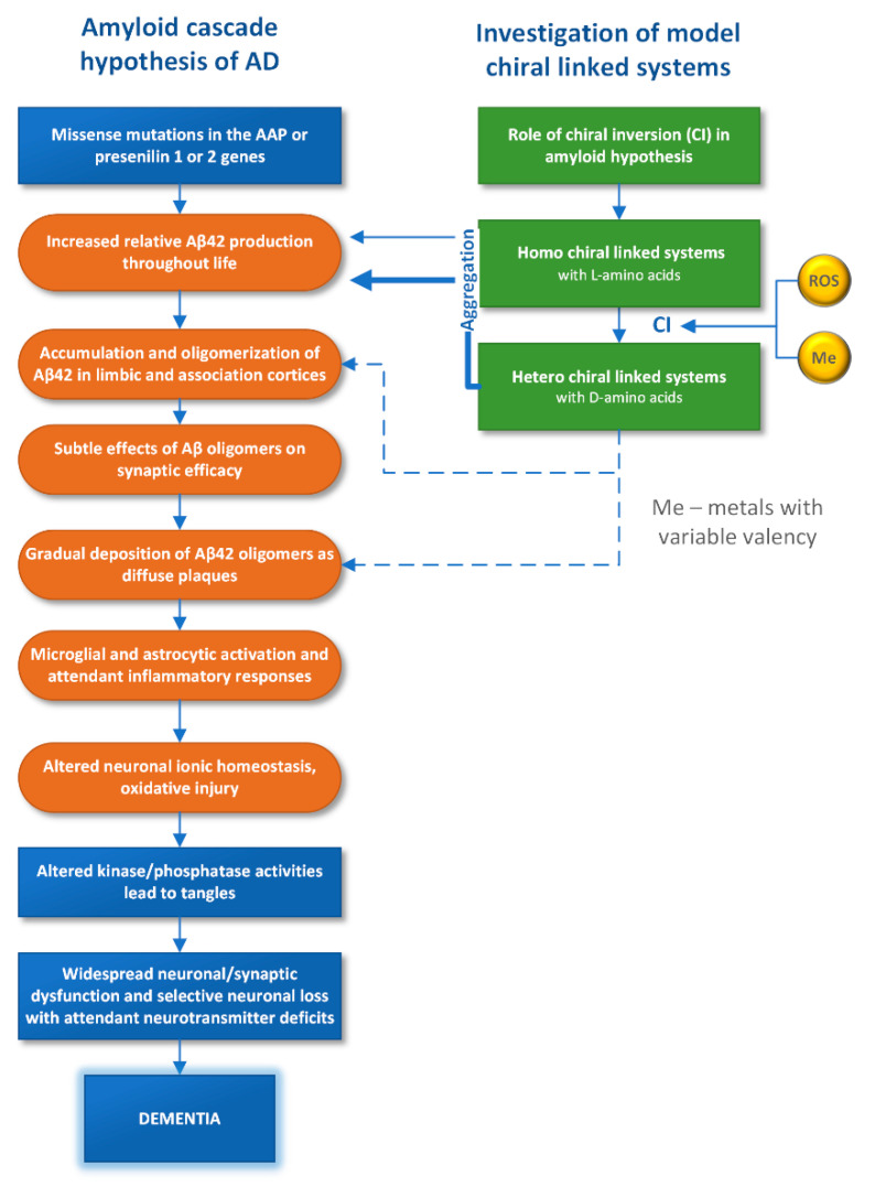
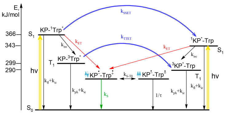
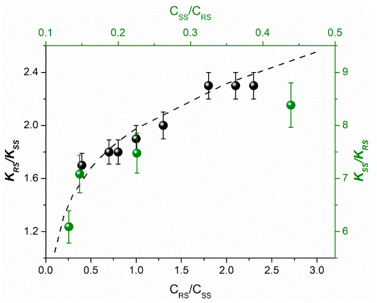
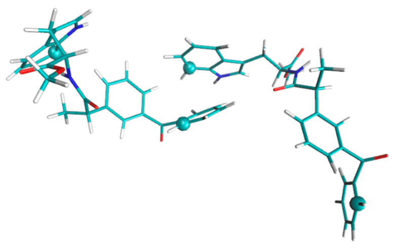
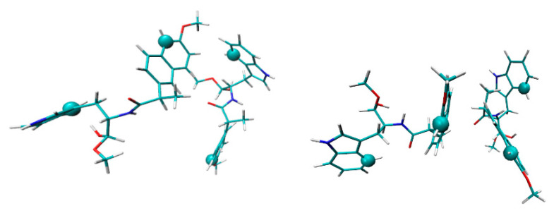
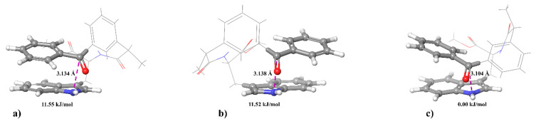
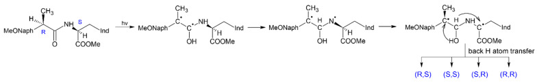
In this review, we provide an illustration of the idea discussed in the literature of using model compounds to study the effect of substitution of L- for D-amino acid residues in amyloid peptides. The need for modeling is due to the inability to study highly disordered peptides by traditional methods (high-field NMR, X-ray). At the same time, the appearance of such peptides, where L-amino acids are partially replaced by D-analogs is one of the main causes of Alzheimer's disease. The review presents examples of the use diastereomers with L-/D-tryptophan in model process-photoinduced electron transfer (ET) for studying differences in reactivity and structure of systems with L- and D-optical isomers. The combined application of spin effects, including those calculated using the original theory, fluorescence techniques and molecular modeling has demonstrated a real difference in the structure and efficiency of ET in diastereomers with L-/D-tryptophan residues. In addition, the review compared the factors governing chiral inversion in model metallopeptides and Aβ42 amyloid.

Keywords: D-amino acids; amyloid peptides; chiral inversion; chiral linked systems; diastereomers; electron transfer; fluorescence quenching; molecular dynamics; spin effects.


Conflict of interest statement

The authors declare no conflict of interest.

Figures

Figure 1
Figure 1
The amyloid cascade hypothesis for Alzheimer’s disease and results of the model systems investigation that provides insight into the role of D-amino acid participation in the amyloid Aβ42 transformation.
Figure 2
Figure 2
Structures of the model linked systems—dyads (IIV).
Scheme 1
Scheme 1
Photoinduced processes in dyad I. Subscripts denote fl—fluorescence, ic—internal conversion, isc—intersystem crossing, ph—phosphorescence, exc—exciplex, S-T0—intersystem crossing in BZ, *—excited states.
Figure 3
Figure 3
The corrected fluorescence emission spectra of KP-Trp dyad compared to the parent N-acetyl-tryptophan methyl ester in acetonitrile, λexc = 280 nm.
Scheme 2
Scheme 2
Photoinduced processes in dyad II. The ratio of light absorbed at λexc = 308 nm by KP/Trp is 55/45, at λexc = 280 nm 65/35. This scheme is similar to the case of dyad I, the difference is that back ET is allowed only from the singlet spin state of BZ. As appears from above, when discussing the CIDNP effects, only the left part of photoinduced processes should be considered.
Figure 4
Figure 4
The experimental dependence of the observed ratios of CIDNP enhancement coefficients and calculated values (dash line) on (R,S)-diastereomer concentration for dyads IV (black balls, CSS = 1 mM) and II (green balls, CSS = 2 mM) [14,20].
Figure 5
Figure 5
The approach of two molecules of (R,S)-diastereomer of dyad II, forming a dimer. The distance between atoms marked by spheres was analyzed [20].
Figure 6
Figure 6
The approach of two molecules of (S,S)-diastereomer of dyad I, forming a different types of dimers: approach of a NPX fragment with Trp (left) and two NPX residues (right). The distance between atoms marked by spheres was analyzed [20].
Figure 7
Figure 7
The optimized structures of (a) (R,S)-; (b) (S,R)-; (c) (S,S)-diastereomers of dyad II. Values of Gibbs energy are given relative to (S,S)-diastereomer [20].
Figure 8
Figure 8
2H-Exchange of ±-flurbiprofenoyl-CoA ester under the action of AMACR that shows change in methyl group multiplet at d 1.45 ppm. (A) Heat-inactivated enzyme; (B) Active enzyme. Reproduced from [29] with permission from The Royal Society of Chemistry.
Figure 9
Figure 9
Proposed mechanism of chiral inversion in the Ni2+-asparagine-cysteine-cysteine metallopeptide.
Figure 10
Figure 10
Scheme of photoinduced CI in (R,S)-configuration of dyad I occurring through the stage of biradical formation and sequential rearrangement in the biradical resulted in formation of a biradical with paramagnetic centers located on pro-chiral centers, where MeONaph is 6-methoxynaphthalen-2-yl, Ind is indol-3-yl, asterisk denotes chiral centers. Adapted from [35], with permission from Elsevier.

Similar articles

Cited by

References

    1. Tverdislov V.A., Yakovenko L.V., Zhavoronkov A.A. Chirality as a problem of biochemical physics. Russ. J. Gen. Chem. 2007;77:1994–2005. doi: 10.1134/S1070363207110291. - DOI
    1. Hardy J., Selkoe D.J. The Amyloid Hypothesis of Alzheimer’s Disease: Progress and Problems on the Road to Therapeutics. Science. 2002;297:353–356. doi: 10.1126/science.1072994. - DOI - PubMed
    1. Raskatov J.A., Teplow D.B. Using chirality to probe the conformational dynamics and assembly of intrinsically disordered amyloid proteins. Sci. Rep. 2017;7:12433. doi: 10.1038/s41598-017-10525-5. - DOI - PMC - PubMed
    1. Chiellini G. Understanding Amyloid Structures and Disease: A Continuing Challenge in Health Research. Int. J. Mol. Sci. 2021;22:6620. doi: 10.3390/ijms22126620. - DOI - PMC - PubMed
    1. Abramov A.Y., Canevari L., Duchen M.R. Calcium signals induced by amyloid PA peptide and their consequences in neurons and astrocytes in culture. Biochim. Biophys. Acta. 2004;1742:81–87. doi: 10.1016/j.bbamcr.2004.09.006. - DOI - PubMed

LinkOut - more resources