Review
. 2022 Mar 12;23(6):3079.
doi: 10.3390/ijms23063079.

Mono a Mano: ZBP1's Love-Hate Relationship with the Kissing Virus

Affiliations
Review

Mono a Mano: ZBP1's Love-Hate Relationship with the Kissing Virus

Alan Herbert et al. Int J Mol Sci. .

Abstract

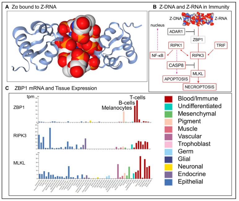
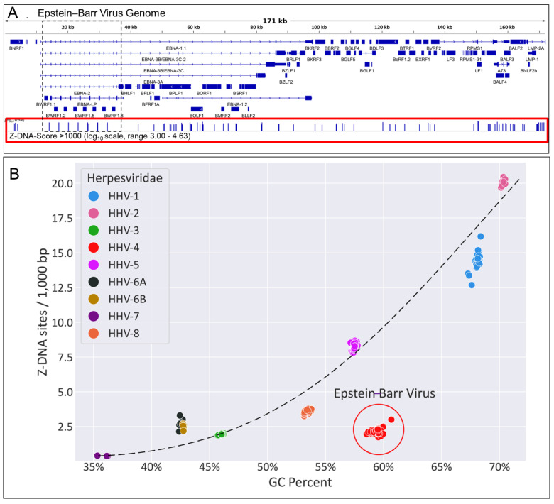
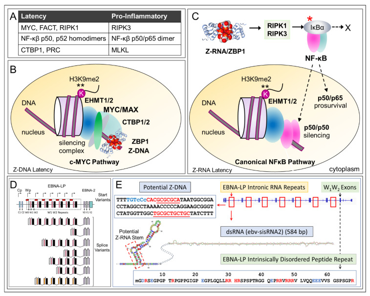
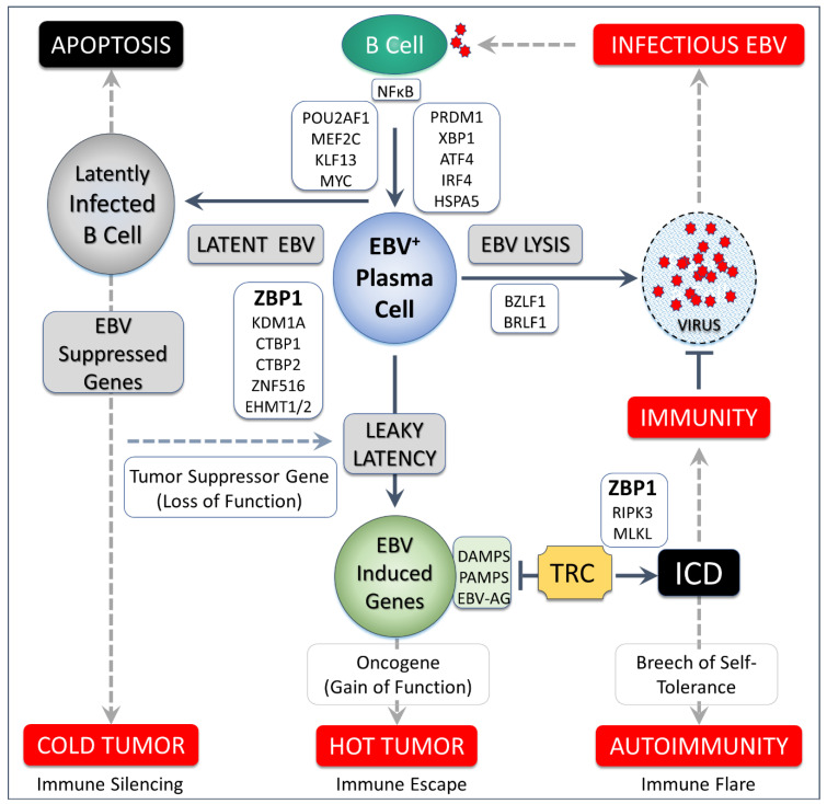
Z-DNA binding protein (ZBP1) very much represents the nuclear option. By initiating inflammatory cell death (ICD), ZBP1 activates host defenses to destroy infectious threats. ZBP1 is also able to induce noninflammatory regulated cell death via apoptosis (RCD). ZBP1 senses the presence of left-handed Z-DNA and Z-RNA (ZNA), including that formed by expression of endogenous retroelements. Viruses such as the Epstein-Barr "kissing virus" inhibit ICD, RCD and other cell death signaling pathways to produce persistent infection. EBV undergoes lytic replication in plasma cells, which maintain detectable levels of basal ZBP1 expression, leading us to suggest a new role for ZBP1 in maintaining EBV latency, one of benefit for both host and virus. We provide an overview of the pathways that are involved in establishing latent infection, including those regulated by MYC and NF-κB. We describe and provide a synthesis of the evidence supporting a role for ZNA in these pathways, highlighting the positive and negative selection of ZNA forming sequences in the EBV genome that underscores the coadaptation of host and virus. Instead of a fight to the death, a state of détente now exists where persistent infection by the virus is tolerated by the host, while disease outcomes such as death, autoimmunity and cancer are minimized. Based on these new insights, we propose actionable therapeutic approaches to unhost EBV.

Keywords: ADAR1; Epstein–Barr virus; Z-DNA; Z-RNA; ZBP1; autoimmune disease; cancer; exhausted T cells; flipons; lymphoma; systemic lupus erythematous.


Conflict of interest statement

Author A.H. is the founder of InsideOutBio, a company that works in the field of immuno-oncology. The remaining authors declare that the research was conducted in the absence of any commercial or financial relationships that could be construed as a potential conflict of interest.

Figures

Figure 1
Figure 1
Z-DNA binding protein 1 (ZBP1) has different roles in development of the immune system. (A). Structure of the Z-DNA binding protein Zα2 domain bound to the zig-zag backbone of left-handed Z-RNA (from PDB:3EY1 rendered by NGL Viewer [22]). (B) ZBP1 senses left-handed Z-DNA and Z-RNA to initiate cell death, either by apoptosis or necroptosis. Both pathways depend on an interaction of receptor interaction protein kinase I and RIPK3. RIPK3 can also be activated by TRIF (toll-like receptor adaptor molecule 1 encoded by TICAM1). Execution of apoptosis depends on caspase 8 activation (CASP8), a protein that also inhibits RIPK3 activation of MLKL( mixed lineage kinase domain-like pseudokinase). ADAR1 (adenosine deaminase RNA specific) inhibits activation of ZBP1 through its Zα domain). RIPK1 is also able to activate the nuclear factor kappa B (NF-κB) that then translocates to the nucleus. (C) Expression of ZBP1 in normal tissues is highest in T cells, B cells and melanocytes as measured by tpm (transcripts per kilobase million).
Figure 2
Figure 2
Single cell analysis of RNA expression in human liver, spleen and blood [23] using the UMAP algorithm implemented in the R-package scater [25] (random seed = 1000). (A). Clusters by cell type. (B) Markers used to assign cell type are listed in the lower left-hand corner (C). Expression of genes relevant to ZBP1 induced necroptosis (ZBP1, RIPK3, MLKL), development (TOX, thymocyte selection associated high mobility group box), proliferation (MKI67, marker of proliferation Ki-67) and tissue residence (CD69). (D). Coexpression of RNAs. The expression level of one gene is indicated by color and that of the other by size. The left panel of each pair is based on two different genes allowing visualization of how often the two genes are coexpressed. The right panel is based on both the size and color of a single gene, giving the maximum coexpression that is possible. (TRAC, T-cell receptor alpha constant region; TRDC, T-cell receptor delta constant region; FGFBP2, fibroblast growth factor binding protein 2). The dataset for this analysis is available in SingleCellExperiment format https://rdrr.io/github/LTLA/scRNAseq/man/ZhaoImmuneLiverData.html (accessed on 7 September 2021) and analyzed using the protocols detailed at https://bioconductor.org/packages/release/bioc/vignettes/scater/inst/doc/overview.html (accessed on 7 September 2021).
Figure 3
Figure 3
A proposed timeline for immune cell development highlighting different roles for ZBP1 in protecting against threats. Early in development, ZBP1 is able to protect against reactivation of endogenous retroelements and aberrant regulation of transcription by sensing the dsRNA involved and inducing apoptosis (indicated by *). Late in development, ZBP1 expressed by tissue-resident immune cells can activate necroptosis (indicated by +) when pathogen associated molecular patterns (PAMPs), damage associated molecular patterns (DAMPs) or live-style associated molecular patterns (LAMPs) are detected. The inflammatory cell death clears space for responding T cells to eliminate the threat. NF-κB is activated by ZBP1 during an adaptive immune response (shown by ♦).
Figure 4
Figure 4
(A). Plasma cells have high expression of unfolded protein response (UPR) genes including key transcriptional factors XBP1 (X-box binding protein 1), IRF4 (interferon regulatory factor 4) and PRDM1 (PR/SET domain 1), along with the regulator HSPA5 (heat shock protein family A (Hsp70) member 5) as shown in the upper panel. The middle panel confirms coexpression with ZBP1 with the smallest dots representing cells that lack ZBP1 expression. (B). Expression of RNA and DNA nucleic acid sensors and their effectors show no or low coexpression in plasma cells relative to other immune cells from blood, liver and spleen (upper two rows). The expression of apoptosis inducing factor (AIFM2) is also low in plasma cells, but the effector proteins PARP1 (poly(ADP-ribose) polymerase 1) and MIF (Macrophage Inhibitory Factor) of the ADP-dependent apoptosis pathway are robustly expressed along with the heat shock 90 protein HASP90B1. (C). Transcription factors expressed in plasma cells belonging to gene families with proposed roles EBV gene expression [45,52]. (D). Coexpression of ZBP1 with the FACT (Facilitates Chromatin Transcription) component SSRP1 (structure specific recognition protein 1) and carboxy terminal binding protein 1 (CTBP1).
Figure 5
Figure 5
ZBP1, flipons and disease with evidence of the evolutionary selection against Z-DNA forming regions in the EBV genome. (A). The EBV genome is characterized by strong Z-DNA forming segments in gene promoters. Vertical lines in the red box show the position of sequences with a Z-Score greater than 1000, as determined using the Z-HUNT3 algorithm [2]. Sequences such as these that change their DNA conformation under physiological conditions are called flipons. They can act as switches to turn gene expression “on” or “off” [71]. The region in the dotted box is expanded on panel D of Figure 6 (B). A plot of GC content of different herpes virus (HHV) genome sequences against the number of sequences per 100,000 base pairs with a high propensity to form Z-DNA under physiological conditions (with ZHUNT3 scores >1000). Independent isolates from the various labeled strains are shown. The dashed line represents the expectation that the number of Z-forming sequences increases with GC content. The Epstein virus group of sequences is shifted to the right of this line, consistent with selection against Z-DNA forming sequences in plasma cells.
Figure 6
Figure 6
Different pathways for ZNA dependent EBV latency. (A). The factors and pathways associated with ZNA latency differ from those shown in Figure 1 that promote ZBP1 induced cell death. The MYC pathways depend on the interaction with the FACT complex as described in the text [72], while the NF-κB dependent transcriptional silencing likely rely on the p50 homodimer in acute infection and the p52 homodimer in chronic infection. Both dimers lack a transactivation domain and are known to induce histone H3K9 methylation [73]. (B). Z-DNA dependent latency. The MYC induced Z-DNA formation localizes ZBP1 and other members of the CTBP1 complex to that region [74]. MYC also binds EHTM1 to induce H3K9 methylation [75]. (C). Z-RNA induced latency. The outcome of NF-κB activation depends on context. In addition to well-known pro-inflammatory roles [76], the p50 homodimer (encoded by NFKB1) can induce H3K9 methylation to suppress interferon induced genes [73]. In this case, phosphorylation of the IκBα (indicated by a red asterisk) protein leads to its ubiquitination and removal by proteolysis (indicated by a cross). The p50 and p65 NF-κB subunits can then enter the nucleus. The non-canonical NF-κB pathway is based on p52 homodimers encoded by NFKB2. (D). The EBV early region contains the essential latency protein EBNA-LP gene (See Figure 5A for entire EBV genome) and repeats of the W1 and W2 exons, as indicated by differently colored vertical stripes. W0, C1, C2 and Wp represent EBV promoters. Alternative transcription start sites are indicated by the bent arrows. RNA splice forms are also presented (based on Figure 1 from [77]). The red boxes indicate the intron repeat sequences that overlap the BWRF1 open reading frame and encode 586 base pair RNA hairpins [64]. (E). The potential EBNA-LP scaffold for regulating EBV gene expression consists of an intronic long-noncoding RNA that forms Z-RNA and RNA hairpins, plus the intrinsically disordered peptide the gene encodes (basic amino acids are colored in red). The Z-DNA forming sequences in the 7 repeats are indicated in red letters with blue indicating an extended Z- forming region that lacks a perfectly alternating purine/pyrimidine repeat (indicated by dotted lines under residues and small letters for the bases out of alternation). The Z-RNA stem (shown within the dotted box) forms from the complementary bases that are underlined. The noncoding RNA and peptide have the potential to nucleate a condensate that functions like Xist to silence the EBV genome.
Figure 7
Figure 7
Epstein–Barr virus persists as a latent infection in B cells. Plasma cell formation can trigger the lytic program with three possible outcomes. The lytic program may complete and produce virus that spreads the infection. Latency may be maintained. Alternatively, suppression of EBV genes may be leaky with variable and partial suppression of viral genes, producing DAMPs (damage associated molecular recognition patterns), PAMPs (pathogen associated molecular recognition patterns) and EBV antigens (EBV-AG) to activate tissue resident cells (TRC) that act as an early warning system and induce inflammatory cell death (ICD). While TRCs may induce immunity, over time autoimmunity may develop. TRCs may also serve as inflammatory drivers of tumorigenesis. White boxes show host encoded transcription factors expressed in plasma cells, some of which regulate activation of EBV lysis [72]. ZBP1 is reported to bind components of the CTBP transcriptional corepressor complex [74] and may target it to Z-DNA formed in EBV promoter regions to maintain latency. EBV induced suppression of tumor suppressor genes and activation of oncogenes, including those encoded by the virus underlie the ~1-1.5% of cancers caused worldwide by the virus.

Similar articles

Cited by

References

    1. Herbert A. Z-DNA and Z-RNA in human disease. Commun. Biol. 2019;2:7. doi: 10.1038/s42003-018-0237-x. - DOI - PMC - PubMed
    1. Ho P.S. Thermogenomics: Thermodynamic-based approaches to genomic analyses of DNA structure. Methods. 2009;47:159–167. doi: 10.1016/j.ymeth.2008.09.007. - DOI - PubMed
    1. Herbert A., Alfken J., Kim Y.G., Mian I.S., Nishikura K., Rich A. A Z-DNA binding domain present in the human editing enzyme, double-stranded RNA adenosine deaminase. Proc. Natl. Acad. Sci. USA. 1997;94:8421–8426. doi: 10.1073/pnas.94.16.8421. - DOI - PMC - PubMed
    1. Herbert A. Mendelian disease caused by variants affecting recognition of Z-DNA and Z-RNA by the Zα domain of the double-stranded RNA editing enzyme ADAR. Eur. J. Hum. Genet. 2020;28:114–117. doi: 10.1038/s41431-019-0458-6. - DOI - PMC - PubMed
    1. Kim S.H., Jung H.J., Lee I.B., Lee N.K., Hong S.C. Sequence-dependent cost for Z-form shapes the torsion-driven B-Z transition via close interplay of Z-DNA and DNA bubble. Nucleic. Acids Res. 2021;49:3651–3660. doi: 10.1093/nar/gkab153. - DOI - PMC - PubMed

LinkOut - more resources