. 2022 Mar 13;23(6):3089.
doi: 10.3390/ijms23063089.

R-Spondin2, a Positive Canonical WNT Signaling Regulator, Controls the Expansion and Differentiation of Distal Lung Epithelial Stem/Progenitor Cells in Mice

Affiliations

R-Spondin2, a Positive Canonical WNT Signaling Regulator, Controls the Expansion and Differentiation of Distal Lung Epithelial Stem/Progenitor Cells in Mice

Ahmed A Raslan et al. Int J Mol Sci. .

Abstract

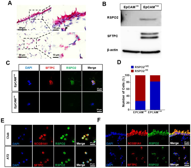
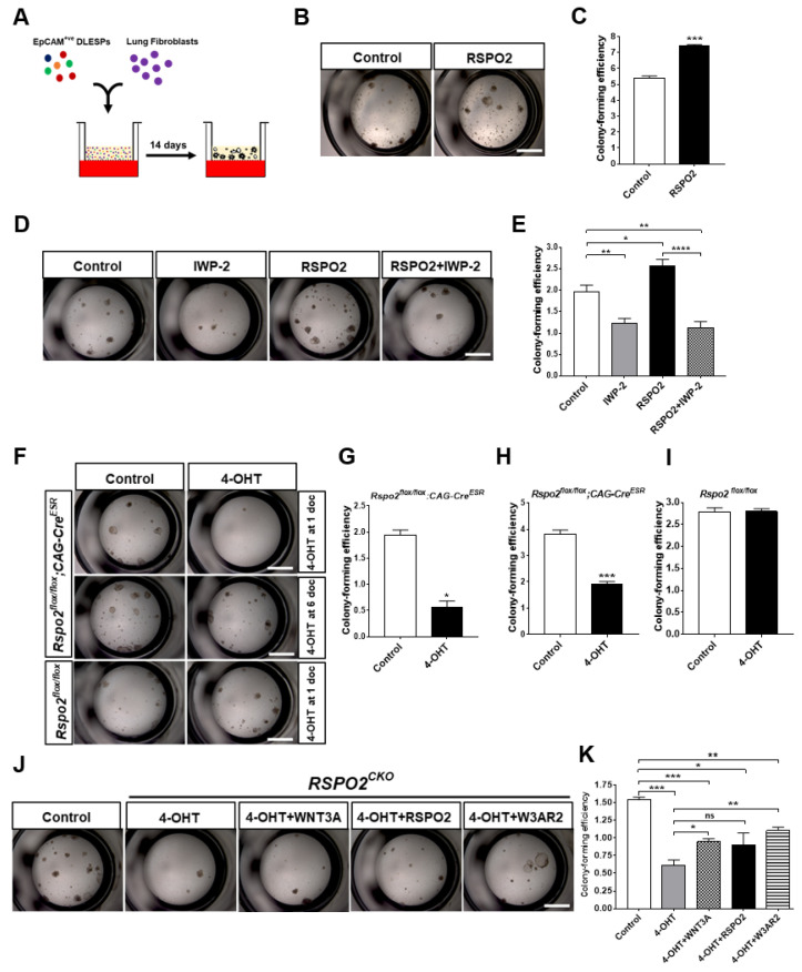
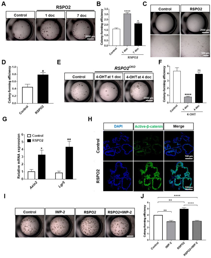
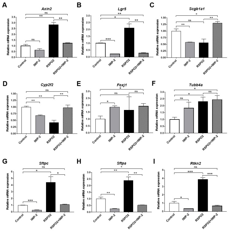
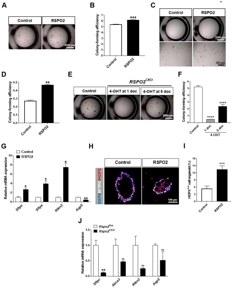
The lungs have a remarkable ability to regenerate damaged tissues caused by acute injury. Many lung diseases, especially chronic lung diseases, are associated with a reduced or disrupted regeneration potential of the lungs. Therefore, understanding the underlying mechanisms of the regenerative capacity of the lungs offers the potential to identify novel therapeutic targets for these diseases. R-spondin2, a co-activator of WNT/β-catenin signaling, plays an important role in embryonic murine lung development. However, the role of Rspo2 in adult lung homeostasis and regeneration remains unknown. The aim of this study is to determine Rspo2 function in distal lung stem/progenitor cells and adult lung regeneration. In this study, we found that robust Rspo2 expression was detected in different epithelial cells, including airway club cells and alveolar type 2 (AT2) cells in the adult lungs. However, Rspo2 expression significantly decreased during the first week after naphthalene-induced airway injury and was restored by day 14 post-injury. In ex vivo 3D organoid culture, recombinant RSPO2 promoted the colony formation and differentiation of both club and AT2 cells through the activation of canonical WNT signaling. In contrast, Rspo2 ablation in club and AT2 cells significantly disrupted their expansion capacity in the ex vivo 3D organoid culture. Furthermore, mice lacking Rspo2 showed significant defects in airway regeneration after naphthalene-induced injury. Our results strongly suggest that RSPO2 plays a key role in the adult lung epithelial stem/progenitor cells during homeostasis and regeneration, and therefore, it may be a potential therapeutic target for chronic lung diseases with reduced regenerative capability.

Keywords: AT2 cells; R-spondin; RSPO2; WNT signaling; club cells; lung homeostasis; lung regeneration; lung stem/progenitor cells; β-catenin.


Conflict of interest statement

The authors declare no conflict of interest.

Figures

Figure 1
Figure 1
RSPO2 is expressed in club and AT2 cells of adult lungs. (A) Single-molecule RNA in situ hybridization detection of Rspo2 RNA. Top and bottom panels represent the airway and alveoli, respectively. The right panels show the magnified images of the dotted rectangular region of the left panels. (B) Western blot analysis of RSPO2 protein expression in freshly isolated EpCAM+ve distal lung epithelial cells. SFTPC and β-actin expression were also determined for validating cell purity and as protein loading control, respectively. (C) Immunofluorescence staining of RSPO2 (green) and AT2 cell marker, SFTPC (red), in freshly isolated EpCAM+ve distal lung epithelial cells. (D) Quantification of RSPO2+ve cell fraction in the isolated distal lung epithelial cells. (E) Co-expression of RSPO2 (green) and club cell marker, SCGB1A1 (red), in the isolated club cells, and RSPO2 (green) and SFTPC (red) in AT2 cells. (F) Immunofluorescence staining of RSPO2 (green), SCGB1A1 (red in the upper panels), and SFTPC (red in the bottom panels) in adult lung tissue section.
Figure 2
Figure 2
Rspo2 is essential for the colony formation of DLESPs in ex vivo 3D organoid culture. (A) Schematic diagram of ex vivo air–liquid interface (ALI) 3D organoid culture. (B,C) Bright-field images and quantification showing the effect of RSPO2 protein treatment (200 ng/mL) on colony formation of DLESPs in the organoid culture for 14 days. (D,E) Bright-field images and colony-forming efficiency of DLESPs showing the effect of WNT protein secretion blocker, IWP-2 (5 μM), in the organoid culture treated for 14 days. (F) Bright-field images of Rspo2CKO DLESP-derived organoids after 14 days of culture. Inactivation of Rspo2 was achieved by 4-OHT (500 nM) treatment for the first two days of organoid culture. (G,H) Colony-forming efficiency of Rspo2CKO DLESPs. The cells were treated with 4-OHT (500 nM) for the first two days (G) or at culture day 6 for two days (H). (I) No 4-OHT effect on colony-forming efficiency of control Rspo2flox/flox DLESPs. 4-OHT treatment was administered for the first two days of the organoid culture and data were collected at day 14. (J,K) Bright-field images and colony-forming efficiency showing the rescued effects of WNT3A (20 ng/mL), RSPO2 (200 ng/mL), or W3AR2 (RSPO2 + WNT3A) protein treatment on Rspo2CKO DLESPs organoid formation. Data are presented as mean ± SEM. ns: no statistical significance, * p < 0.05, ** p < 0.01, *** p < 0.001, **** p < 0.0001.
Figure 3
Figure 3
RSPO2 supports the colony formation of club cells in ex vivo 3D organoid culture through the activation of endogenous WNT/β-catenin signaling. (A,B) Bright-field images and colony-forming efficiency of club cells in the 3D organoid culture showing the effect of RSPO2 treatment (200 ng/mL). RSPO2 was administered from day 1 of culture (1 doc) or day 7 of organoid culture (7 doc). (C,D) Bright-field images and colony-forming efficiency of club cells in a fibroblast-free, FGF10/HGF supplemented 3D organoid culture. RSPO2 (200 ng/mL) was added from 1 doc for 7 days. (E,F) Bright-field images and colony-forming efficiency of Rspo2CKO club cells in the organoid culture. (G) RSPO2 (200 ng/mL) upregulated the expression of two WNT/β-Catenin downstream target genes, Axin2 and Lgr5, in club cell-derived organoids, as determined by qRT-PCR analysis. (H) Immunofluorescence staining of active β-Catenin (unphosphorylated form) after RSPO2 treatment in club cell-derived organoids. Cell nuclei were counterstained with DAPI. (I,J) Bright-field images and colony-forming efficiency of club cells showing the effect of WNT protein secretion blocker, IWP-2 (5 μM), on the organoid culture (Scale bar, 2000 μm). All organoids were harvested at 7 or 14 doc for further analysis. Data are presented as mean ± SEM. ns: no statistical significance, * p < 0.05, ** p < 0.01, **** p < 0.0001.
Figure 4
Figure 4
RSPO2 promotes the differentiation of club cells into multiple epithelial lineages in ex vivo 3D organoid culture. (A) qRT-PCR analysis of club cell marker genes, Scgb1a1, Cyp2f2 and Bpifa1, in club cell-derived organoids treated with RSPO2 (200 ng/mL) for 14 days. (B) qRT-PCR analysis of gene markers for ciliated (Foxj1), basal (Krt5), and goblet (Muc5ac) cells in the RSPO2 (200 ng/mL)-treated club cell-derived organoids. (C,D) qRT-PCR analysis of AT2 cell marker genes (C) Sftpc, Abca3, and Sftpa, and AT1 cell marker genes (D) Rtkn2, Aqp5, and Pdpn in the RSPO2-treated club cell-derived organoids. (E) qRT-PCR analysis of various differentiated cell marker genes in Rspo2CKO club cell-derived organoids. (F,G) Immunofluorescence staining images for SCGB1A1, FOXJ1, SFTPC, and PDPN, and quantification of SCGB1A1+ve club, FOXJ1+ve ciliated, and SFTPC+ve AT2 cells in the RSPO2 (200 ng/ml)-treated club cell organoids. All organoids were collected after 14 days in culture. Data are presented as mean ± SEM. ns: no statistical significance, * p < 0.05, ** p < 0.01, **** p < 0.0001.
Figure 5
Figure 5
RSPO2 drives the differentiation of club cells into alveolar lineages in an endogenous WNT/β-catenin activation-dependent manner. (A,B) qRT-PCR analysis of WNT/β-catenin downstream target genes, Axin2 and Lgr5, in IWP-2 (5 μM)- and/or RSPO2 (200 ng/mL)-treated club cell-derived organoids. (CI) qRT-PCR analysis of club cell markers Scgb1a1 (C) and Cyp2f2 (D), ciliated cell markers Foxj1 (E) and Tubb4a (F), AT2 cell markers Sftpc (G) and Sftpa (H), and AT1 cell marker Rtkn2 (I) in the club cell organoids treated with IWP-2, RSPO2, or both. All organoids were collected for RNA isolation at day 14 of culture. Data are presented as mean ± SEM. ns: no statistical significance, * p < 0.05, ** p < 0.01, *** p < 0.001.
Figure 6
Figure 6
RSPO2 is essential for the colony formation and differentiation of AT2 cells in ex vivo 3D organoid culture. (A,B) Bright-field images and colony-forming efficiency of AT2 cells in ex vivo 3D organoid culture treated with in RSPO2 (200 ng/mL). (C,D) Bright-field images and quantification of colony forming efficiency of AT2 cells in a fibroblast-free, FGF10/HGF supplemented 3D organoid culture in the presence of RSPO2 (200 ng/mL). (E,F) Bright-field images and colony-forming efficiency of Rspo2CKO AT2 cells in the organoid culture. 4-OHT (500 nM) was administered for a total of two days beginning at day 1 or day 5 of organoid culture. (G) qRT-PCR analysis of AT2 cell markers, Sftpc and Sftpa, and AT1 cell markers, Rtkn2 and Aqp5, in AT2 cell-derived organoids treated with RSPO2 (200 ng/mL). (H) Immunofluorescence staining of SFTPC and early AT1 cell marker, HOPX, in AT2 cell-derived organoids treated with RSPO2 (200 ng/mL). DAPI was used to counterstain cell nuclei. (I) Quantification of HOPX+ve cells in the RSPO2-treated AT2 cell organoids. (J) Expression of Sftpc, Abca3, Rtkn2, and Aqp5 in Rspo2CKO AT2 cell organoids by qRT-PCR. All organoids were collected at 7 doc for analysis. Data are presented as mean ± SEM. ns: no statistical significance, * p < 0.05, ** p < 0.01, *** p < 0.001, **** p < 0.0001.
Figure 7
Figure 7
Rspo2 gene ablation causes defects in airway regeneration after naphthalene-induced acute lung injury. (A) Rspo2 expression during airway regeneration in naphthalene-induced lung injury by qRT-PCR. dai; days after injury. (B) Immunofluorescence staining of RSPO2 and SCGB1A1 during airway regeneration. (C) Schematic diagram of the experimental schedule for conditional Rspo2 knockout followed by naphthalene-induced acute lung injury, and the expression of SCGB1A1 and Ac-α-tubulin determined by immunofluorescence staining of Rspo2CKO lung tissues at 14 dai. (D) Measurement of the thickness of the bronchiolar epithelium at 14 dai in Rspo2CKO lungs. (E,F) The percentage of SCGB1A1+ve club and Ac-α-tubulin+ve ciliated cells in the bronchiolar epithelium at 14 dai in Rspo2CKO lungs. (G,H) Immunofluorescence staining of Ki67 and active β-Catenin at 14 dai in Rspo2CKO lung tissue and quantification of Ki67+ve proliferative cells. Data were collected from the samples (n = 4 for Rspo2CKO and control mice each) and are presented as mean ± SEM. ns: no statistical significance, * p < 0.05, ** p < 0.01.

Similar articles

Cited by

References

    1. Raslan A.A., Yoon J.K. WNT Signaling in Lung Repair and Regeneration. Mol. Cells. 2020;43:774–783. doi: 10.14348/molcells.2020.0059. - DOI - PMC - PubMed
    1. Wansleeben C., Barkauskas C.E., Rock J.R., Hogan B.L. Stem cells of the adult lung: Their development and role in homeostasis, regeneration, and disease. Wiley Interdiscip. Rev. Dev. Biol. 2013;2:131–148. doi: 10.1002/wdev.58. - DOI - PubMed
    1. Lee J.H., Rawlins E.L. Developmental mechanisms and adult stem cells for therapeutic lung regeneration. Dev. Biol. 2018;433:166–176. doi: 10.1016/j.ydbio.2017.09.016. - DOI - PubMed
    1. Hogan B.L., Barkauskas C.E., Chapman H.A., Epstein J.A., Jain R., Hsia C.C., Niklason L., Calle E., Le A., Randell S.H., et al. Repair and regeneration of the respiratory system: Complexity, plasticity, and mechanisms of lung stem cell function. Cell Stem Cell. 2014;15:123–138. doi: 10.1016/j.stem.2014.07.012. - DOI - PMC - PubMed
    1. Lynch T.J., Anderson P.J., Rotti P.G., Tyler S.R., Crooke A.K., Choi S.H., Montoro D.T., Silverman C.L., Shahin W., Zhao R., et al. Submucosal Gland Myoepithelial Cells Are Reserve Stem Cells That Can Regenerate Mouse Tracheal Epithelium. Cell Stem Cell. 2018;22:653–667.E5. doi: 10.1016/j.stem.2018.03.017. - DOI - PMC - PubMed

LinkOut - more resources