. 2022 Mar 13;23(6):3095.
doi: 10.3390/ijms23063095.

A Homozygous Deep Intronic Variant Causes Von Willebrand Factor Deficiency and Lack of Endothelial-Specific Secretory Organelles, Weibel-Palade Bodies

Affiliations

A Homozygous Deep Intronic Variant Causes Von Willebrand Factor Deficiency and Lack of Endothelial-Specific Secretory Organelles, Weibel-Palade Bodies

Hamideh Yadegari et al. Int J Mol Sci. .

Abstract

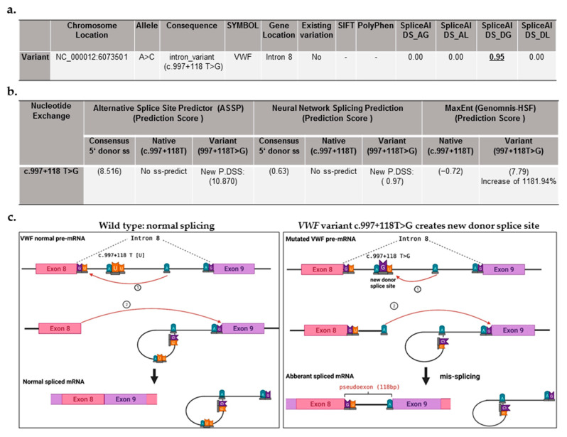
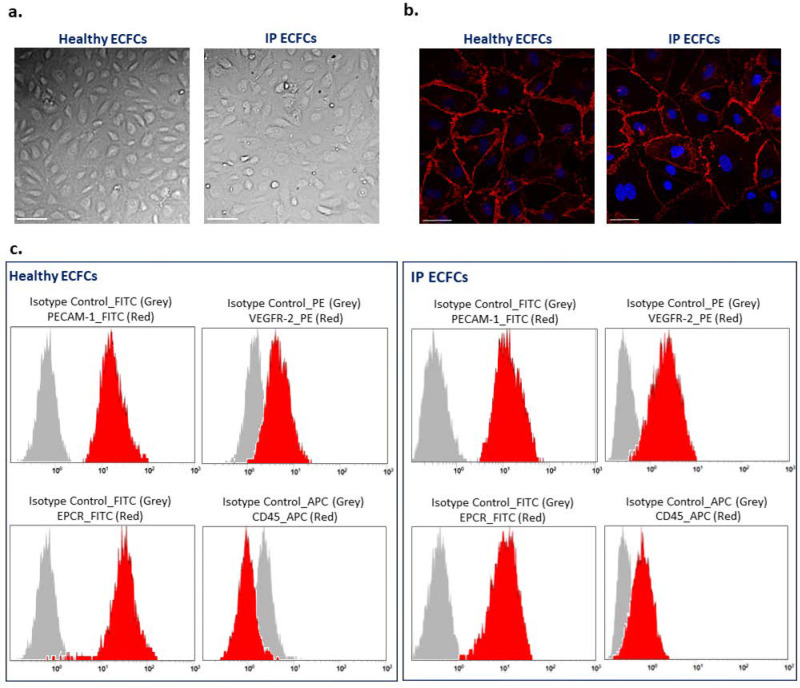
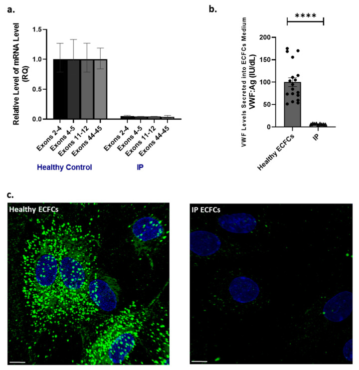
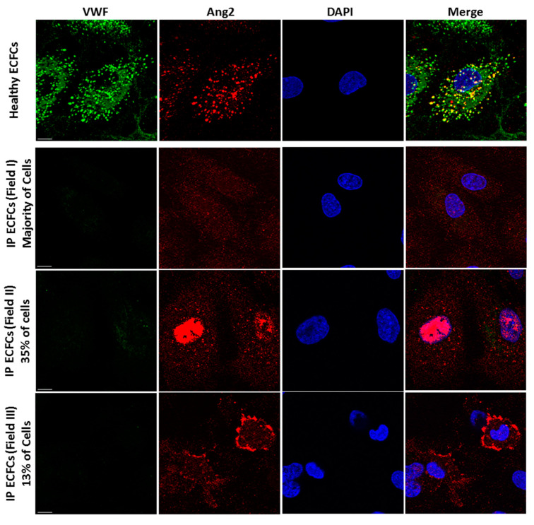
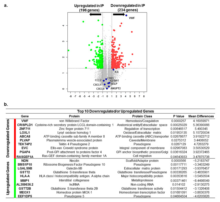
A type 3 von Willebrand disease (VWD) index patient (IP) remains mutation-negative after completion of the conventional diagnostic analysis, including multiplex ligation-dependent probe amplification and sequencing of the promoter, exons, and flanking intronic regions of the VWF gene (VWF). In this study, we intended to elucidate causative mutation through next-generation sequencing (NGS) of the whole VWF (including complete intronic region), mRNA analysis, and study of the patient-derived endothelial colony-forming cells (ECFCs). The NGS revealed a variant in the intronic region of VWF (997 + 118 T > G in intron 8), for the first time. The bioinformatics assessments (e.g., SpliceAl) predicted this variant creates a new donor splice site (ss), which could outcompete the consensus 5′ donor ss at exon/intron 8. This would lead to an aberrant mRNA that contains a premature stop codon, targeting it to nonsense-mediated mRNA decay. The subsequent quantitative real-time PCR confirmed the virtual absence of VWF mRNA in IP ECFCs. Additionally, the IP ECFCs demonstrated a considerable reduction in VWF secretion (~6% of healthy donors), and they were devoid of endothelial-specific secretory organelles, Weibel−Palade bodies. Our findings underline the potential of NGS in conjunction with RNA analysis and patient-derived cell studies for genetic diagnosis of mutation-negative type 3 VWD patients.

Keywords: ECFCs; Weibel–Palade bodies; angiopoietin-2; deep intronic mutation; next-generation sequencing; von Willebrand disease; von Willebrand factor.


Conflict of interest statement

The authors declare no conflict of interest.

Figures

Figure 1
Figure 1
Detection of a deep intronic variant in von Willebrand factor (VWF) gene after executing next-generation sequencing (NGS) and subsequent bioinformatic analysis. (a) Data obtained from analysis of variant c.997 + 118 T > G, detected by NGS, using Ensemble Variant Effect Predictor (VEP), including variant annotation, variant location, the existing variant in public variant databases, and predicting its pathogenic effect. Genomic location of VWF variant is annotated based on latest assembly (GRCh38/hg38: chr12: 5,948,877–6,124,770). No: not existing in the public variant databases. SpliceAl (Illumina artificial intelligence splicing prediction software) prediction scores include DS_AG (delta score for acceptor gain), DS_AL (delta score for acceptor loss), DS_DG (delta score for donor gain), and DS_DL (delta score for donor loss). The calculated scores range from 0 to 1 and can be interpreted as the probability of the variant being splice-altering. The suggested cutoffs are 0.2 (high recall), 0.5 (recommended), and 0.8 (high precision). The VEP-SpliceAl tool predicted the variant c.997 + 118 T > G induces the gain of a donor splice site, providing the DS_DG score of 0.95. -, not applicable, the SIFT and Polyphen predict the effect of exonic variants on protein function/structure. (b) Summary of bioinformatic analysis (by Neural Network Splicing, Alternative Splice Site Predictor, and plug-in MaxEnt of the Human Splicing Finder (HSF)), predicting the impact of the VWF gene (VWF) variant c.997 + 118 T > G on splicing processing. ss, splicing site; P.DSS, potential donor splice site. (c) Schematic image of normal splicing of the wild-type VWF mRNA at exon/intron 8 junction (left side) as well as the mis-splicing due to the variant c.997 + 118 T > G, embedded in intron 8 of VWF, which creates a new donor splice site, leading to an aberrant mRNA with exon elongation (a pseudoexon of 118 base pairs (bp)).
Figure 2
Figure 2
Phenotype characterization of endothelial colony-forming cells (ECFCs). (a) Typical endothelial cobblestone-like morphology of ECFCs isolated from a healthy individual and the index patient (IP) observed using bright-field microscopy. Scale bars, 100 µm. (b) Expression of VE-cadherin (red) at cell–cell junctions is visualized with immunostaining of ECFCs derived from a healthy individual (left side) and patient (IP ECFCs) (right side). The nucleus is stained with DAPI (blue). Scale bars, 50 µm. (c) Flow cytometry analysis of ECFCs derived from a healthy donor (left images) and the IP (right images). The data confirmed that both healthy and IP isolated cells were positive for endothelial cell canonical markers of PECAM-1 (CD31-FITC conjugated), VEGFR-2 (PE-conjugate), and EPCR (FITC-conjugate); besides, they were negative for leukocyte cell marker CD45 (APC-conjugate). Isotype controls were conjugated with either FITC, PE, or APC corresponding to their relevant antibodies, and they are shown as grey bell curve graphs.
Figure 3
Figure 3
Von Willebrand factor (VWF) mRNA expression, as well as VWF secretion and intracellular storage, in endothelial colony-forming cells (ECFCs). (a) Illustration of comparative VWF mRNA levels in the index patient (IP) ECFCs quantified by real-time PCR, using primer/probe combinations directing four different sites in VWF cDNA, across exons 2–4, 4–5, 11–12, and 43–45. The measurements were performed based on the comparative CT(∆∆ CT) method. Measurements of VWF mRNA levels were normalized to endogenous glyceraldehyde-3-phosphate dehydrogenase (GAPDH) or actin beta (ACTB) mRNA. (b) Graph of the mean of VWF antigen (VWF:Ag) levels in the medium of ECFCs obtained from IP (three independent ECFC isolations, three different passages each; N = 9) and six healthy donors (three different passages each; N = 18). (c) Immunofluorescence images of ECFCs isolated from the IP and healthy individuals. In healthy ECFCs, VWF (green) is deposited in stick-shaped organelles, resembling Weibel–Palade bodies (WPBs). However, VWF staining in the IP-ECFCs showed almost a lack of WPB formation, except for a few WPBs (1 to 3) in some cells. The scale bar is 10 µm.
Figure 4
Figure 4
Altered trafficking of angiopoietin-2 (Ang2), the inflammatory cargo of Weibel–Palade bodies (WPBs), in index patient (IP) endothelial colony-forming cells (ECFCs). Storage of VWF (green) and Ang2 (red) in WPBs is visualized by immunofluorescence staining and subsequent microscopy analysis (by Zeiss Apotome.2 microscopy) of normal ECFCs (upper section). The merge of green and red channels displays the colocalization of VWF with Ang2 in healthy ECFCs. In the IP ECFCs, in absence of VWF and WPBs, the storage of Ang2 is changed. In the majority of IP ECFCs, the Ang2 is spread throughout the cytoplasm (the second row of images); in about 35% of IP cells, the Ang2 is accumulated nearby the nucleus (third row); and in about 13% of IP cells, the Ang2 signals are enriched at the cell periphery (lower row images). Scale bars, 10 µm.
Figure 5
Figure 5
Differentially expressed genes (DEGs) and the top 10 downregulated or upregulated genes in index patient (IP) endothelial colony-forming cells (ECFCs). (a) Volcano plot demonstrates significantly DEGs in IP ECFCs in which –log10 (p) is plotted against the mean differences. Green dots represent the upregulated genes and red dots represent downregulated genes. Numbers of genes upregulated or downregulated are indicated. Dashed lines represent the threshold of statistical significance. DEGs were considered significant when p-value < 0.05 and fold change was greater than 2 with respect to healthy ECFC samples or absolute log2FC (log2 fold change or called mean difference) was more than 1. A negative mean difference value points to lower expression in healthy controls (upregulated expression in IP), and a positive mean difference indicates lower expression in IP-ECFCs. The blue dots illustrate the expression of the VWF, the main Weibel–Palade body (WPB) cargo, which is significantly downregulated, as well as expression of inflammatory cargos of WPBs (Ang2, CCL2, CD63, IGFBP7, CXCL8, IL6, CXCL1, and SELP), which does not show any changes compared with healthy ECFCs. Ang2 (ANGP2), angiopoietin-2; CCL2 (known as MCP-1), C-C motif chemokine ligand 2; IGFBP7, insulin-like growth factor binding protein 7; CXCL8 (known as interleukin 8 (IL8)), C-X-C motif chemokine ligand 8; IL6, interleukin 6; CXCL1 (knows as GROa), C-X-C motif chemokine ligand 1; SELP, P-selectin. (b) The table presents the top ten downregulated as well as top ten upregulated genes in the ECFCs isolated from the patient. The list is generated according to the mean difference (absolute log2FC), and they are sorted by decreasing p-value in the present table.

Similar articles

Cited by

References

    1. Lenting P.J., Christophe O.D., Denis C.V. Von Willebrand factor biosynthesis, secretion, and clearance: Connecting the far ends. Blood. 2015;125:2019–2028. doi: 10.1182/blood-2014-06-528406. - DOI - PubMed
    1. Leebeek F.W.G., Eikenboom J.C.J. Von Willebrand’s Disease. N. Engl. J. Med. 2017;376:701–702. doi: 10.1056/NEJMra1601561. - DOI - PubMed
    1. Lillicrap D. Von Willebrand disease: Advances in pathogenetic understanding, diagnosis, and therapy. Blood. 2013;122:3735–3740. doi: 10.1182/blood-2013-06-498303. - DOI - PMC - PubMed
    1. Castaman G. How I treat von Willebrand disease. Thromb. Res. 2020;196:618–625. doi: 10.1016/j.thromres.2020.07.051. - DOI - PubMed
    1. James P.D., Connell N.T., Ameer B., Di Paola J., Eikenboom J., Giraud N., Haberichter S., Jacobs-Pratt V., Konkle B., McLintock C., et al. ASH ISTH NHF WFH 2021 guidelines on the diagnosis of von Willebrand disease. Blood Adv. 2021;5:280–300. doi: 10.1182/bloodadvances.2020003265. - DOI - PMC - PubMed