. 2022 Mar 14;23(6):3125.
doi: 10.3390/ijms23063125.

A Novel Mechanism of Macrophage Activation by the Natural Yolkin Polypeptide Complex from Egg Yolk

Affiliations

A Novel Mechanism of Macrophage Activation by the Natural Yolkin Polypeptide Complex from Egg Yolk

Wioletta Kazana et al. Int J Mol Sci. .

Abstract

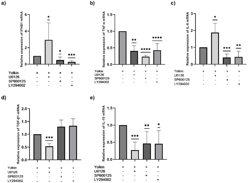
Ageing is accompanied by the inevitable changes in the function of the immune system. It provides increased susceptibility to chronic infections that have a negative impact on the quality of life of older people. Therefore, rejuvenating the aged immunity has become an important research and therapeutic goal. Yolkin, a polypeptide complex isolated from hen egg yolks, possesses immunoregulatory and neuroprotective activity. Considering that macrophages play a key role in pathogen recognition and antigen presentation, we evaluated the impact of yolkin on the phenotype and function of mouse bone marrow-derived macrophages of the BMDM cell line. We determined yolkin bioavailability and the surface co-expression of CD80/CD86 using flow cytometry and IL-6, IL-10, TGF-β and iNOS mRNA expression via real-time PCR. Additionally, the impact of yolkin on the regulation of cytokine expression by MAPK and PI3K/Akt kinases was determined. The stimulation of cells with yolkin induced significant changes in cell morphology and an increase in CD80/CD86 expression. Using pharmaceutical inhibitors of ERK, JNK and PI3K/Akt, we have shown that yolkin is able to activate these kinases to control cytokine mRNA expression. Our results suggest that yolkin is a good regulator of macrophage activity, priming mainly the M1 phenotype. Therefore, it is believed that yolkin possesses significant therapeutic potential and represents a promising possibility for the development of novel immunomodulatory medicine.

Keywords: immunomodulators; innate immunity; macrophage polarization; macrophages; yolkin.


Conflict of interest statement

The authors declare no conflict of interest.

Figures

Figure 1
Figure 1
Analysis of yolkin uptake by BMDM cells over time. The cell membrane was stained with red fluorescent cell linker PKH26 prior to seeding on the tissue culture plate (3 × 10⁵/mL). Next, the cells were stimulated with labelled yolkin (100 µg/mL) for 15, 30 and 60 min and then analyzed using FACS. The quantity (%) of double-positive cells (PKH26 + YOLKIN) over time was measured. The results represent three independent experiments with representative flow cytometry dot plots (a) and a graph of changes over time, presented as median with SD (b). The post-measured analysis was performed using FlowJo VX.07 software, and a one-sample t-test was used to examine the mean differences between actual mean and theoretical mean (0), all with ** p ≤ 0.005, and **** p ≤ 0.0001.
Figure 2
Figure 2
Effect of yolkin on viability and proliferation of bone marrow-derived macrophages (BMDM). The cells (1 × 10⁵/mL) were seeded in 96-well plates in DMEM + 10% FBS and incubated overnight at 37 °C. Next, the cells were exposed to yolkin (10, 100 and 150 µg/mL) for 24 and 48 h. The cell proliferation activity of yolkin was assessed with an MTT assay. Non-stimulated cells were used as a negative control. The results represent three to five independent experiments and data are presented as median ± min–max. A one-sample t-test was used to examine the mean differences between samples and control (100). * p ≤ 0.05, *** p ≤ 0.0001.
Figure 3
Figure 3
Morphology of yolkin- or LPS-stimulated BMDM cells. Stimulation of BMDM cells for 24 or 48 h with yolkin (10 and 100 µg/mL) or LPS (1 µg/mL) induced morphological changes that were visible under a light microscope with 4× lenses. The cells in the control group showed a round-shaped morphology, which changed after exposure to LPS or yolkin, where more spindle-shaped cells were presented. The cells were examined via light microscopy (Leica DM FRE2, bright field). One representative experiment is shown in this figure. Scale bars represent 500 microns.
Figure 4
Figure 4
Analysis of co-expression of CD80 and CD86 on BMDM cells. The cells (3 × 10⁵/mL) were stimulated with yolkin (100µg/mL) or LPS (1 µg/mL) for 24 h and then stained with fluorescent CD80 (1:100) and CD86 (1:100) mouse antibodies. The cells were analyzed using FACS. The results represent three independent experiments with representative flow cytometry dot plots (a) and a graph summarizing the changes, presented as median with SD (b). The cells stimulated with yolkin or LPS were compared with the non-stimulated control cells. The post-measurement analysis was performed using FlowJo VX.07 software and data were analyzed with a one-way ANOVA (Brown–Forsythe and Welch’s test). ** p ≤ 0.005; **** p ≤ 0.0001.
Figure 5
Figure 5
The impact of yolkin on the regulation of iNOS (a), IL-6 (b), IL-10 (c) and TGFβ (d) mRNA expression. BMDM cells (1 × 10⁶/mL) were stimulated with yolkin (100 μg/mL) or LPS (1 μg/mL). After 4 h of stimulation, total RNA was isolated from the cells and the expression of iNOS (a), IL-6 (b), IL-10 (c) and TGFβ (d) mRNA was determined using real-time PCR tests. Experiments were repeated at least three times and data are presented in relative expression units, where Actb was used to normalize all samples. Control cells were assigned an arbitrary value of 1. A one-sample t-test was used to examine the mean differences between samples and control; * p ≤ 0.05; *** p ≤ 0.001 vs. control.
Figure 6
Figure 6
The impact of yolkin on IL-6 (a) and IL-10 (b) production by BMDM cells. BMDM cells (1 × 10⁶/mL) were stimulated with yolkin (10 and 100 ug/mL) or LPS (1 μg/mL). After 24 h of stimulation, supernatants were collected, and the level of cytokines was determined using ELISA. Results are presented as mean with min–max (n = 3–4). A Kruskal–Wallis test was used to examine the mean differences between samples and control * p ≤ 0.05; **** p ≤ 0.00001; ns (not significant) vs. control.
Figure 7
Figure 7
MAPK and PI3K/Akt-dependent regulation of cytokine expression after yolkin treatment of BMDM cells. BMDM cells (1 × 10⁶/mL) were pre-incubated for 1 h with selective kinase inhibitors: 20 μM U0126 (ERK1/2), 25 μM SP600125 (JNK) or 20 μM LY294002 (PI3K/Akt). Next, the cells were stimulated with yolkin (100 μg/mL) for 4 h. After stimulation, total RNA was isolated from the cells, and the expression of IFNβ1 (a), TNF-α (b), IL-6 (c), TGFβ (d) and IL-10 (e) mRNA was determined via real-time PCR. Experiments were repeated at least three times and data are presented in relative expression units, where Actb was used to normalize all samples. Yolkin-treated cells were assigned an arbitrary value of 1. Results are presented as mean ± SD. A one-sample t-test was used to examine the mean differences between yolkin-treated samples and yolkin with inhibitor-treated groups; * p ≤ 0.05, ** p ≤ 0.001, *** p ≤ 0.0001; **** p ≤ 0.00001; vs. yolkin.
Figure 8
Figure 8
Recognition of yolkin samples by innate immune receptors. HEK BlueTM cells were seeded on the plate and stimulated with yolkin samples in 3 repetitions and at concentrations of 10 µg/mL and 100 µg/mL according to the manufacturer’s instruction. To analyze yolkin recognition over time, a set of absorbance measurements was performed at 610, 630 and 650 nm. Then, 630 nm was selected as the representative. LPS was used as a positive control for TLR4 cells and negative for TLR2 and NOD2 cell lines. A positive control for TLR2 recognition was PAMP and for the NOD2 receptor it was MDP. All of the obtained results were statistically significant. A one-way ANOVA with Dunnett’s multiple comparison test was used to compare treated cells with non-treated control (medium). **** p ≤ 0.0001.

Similar articles

Cited by

References

    1. Mine Y., Kovacs-Nolan J. New Insights in Biologically Active Proteins and Peptides Derived from Hen Egg. World’s Poult. Sci. J. 2006;62:87–96. doi: 10.1079/WPS200586. - DOI
    1. Zambrowicz A., Dąbrowska A., Bobak Ł., Szołtysik M. Egg Yolk Proteins and Peptides with Biological Activity. Postepy. Hig. Med. Dosw. 2014;68:1524–1529. doi: 10.5604/17322693.1133600. - DOI - PubMed
    1. Li H., Zhang S. Functions of Vitellogenin in Eggs. Results Probl. Cell Differ. 2017;63:389–401. doi: 10.1007/978-3-319-60855-6_17. - DOI - PubMed
    1. Polanowski A., Sosnowska A., Zabłocka A., Janusz M., Trziszka T. Immunologically Active Peptides That Accompany Hen Egg Yolk Immunoglobulin Y: Separation and Identification. Biol. Chem. 2013;394:879–887. doi: 10.1515/hsz-2012-0337. - DOI - PubMed
    1. Polanowski A., Zabłocka A., Sosnowska A., Janusz M., Trziszka T. Immunomodulatory Activity Accompanying Chicken Egg Yolk Immunoglobulin Y. Poult. Sci. 2012;91:3091–3096. doi: 10.3382/ps.2012-02546. - DOI - PMC - PubMed