Review
. 2022 Mar 15;23(6):3132.
doi: 10.3390/ijms23063132.

Unraveling the Role of Sex Hormones on Keratinocyte Functions in Human Inflammatory Skin Diseases

Affiliations
Review

Unraveling the Role of Sex Hormones on Keratinocyte Functions in Human Inflammatory Skin Diseases

Rossella Gratton et al. Int J Mol Sci. .

Abstract

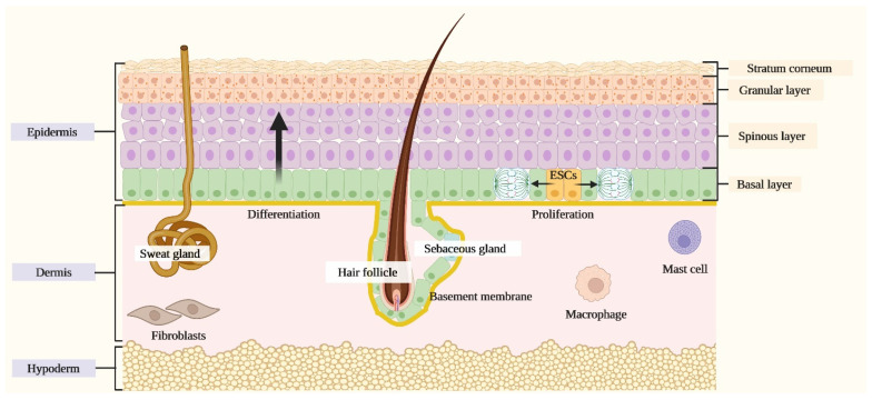
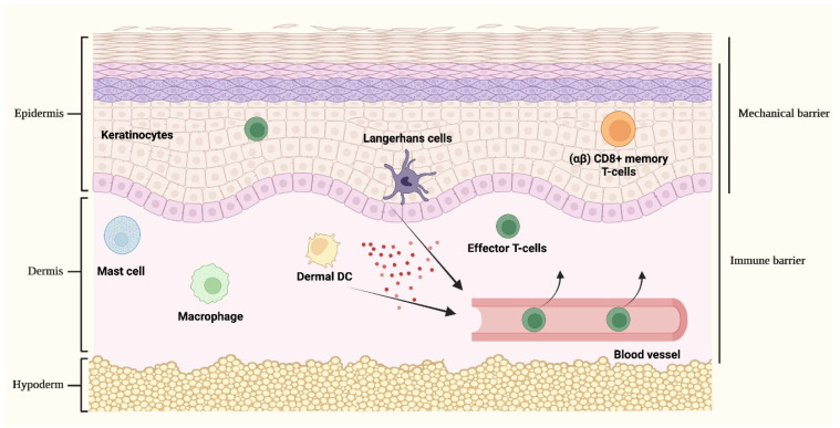
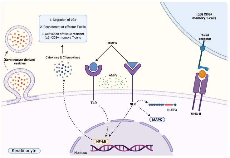
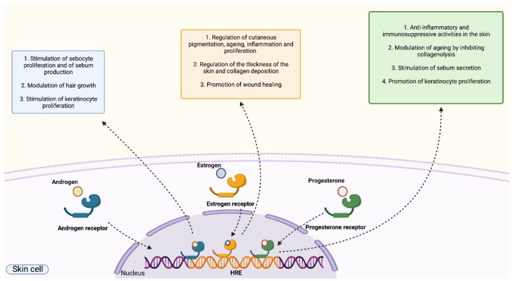
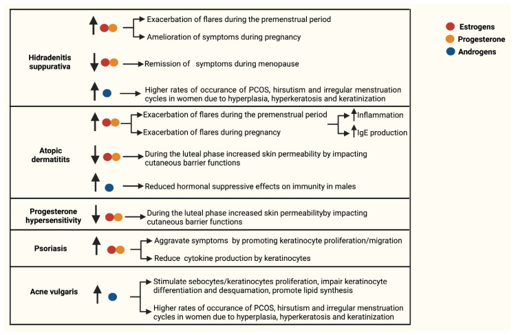
The skin exerts several fundamental functions that are the first physical, chemical and immune barriers to the human body. Keratinocytes, the main cell type of the epidermis, provide mechanical defense, support skin integrity and actively endorse cutaneous immune responses. Not surprisingly, considering these crucial activities, alterations in keratinocyte functions are associated with different inflammatory skin diseases. Recent findings indicate that the skin should not only be regarded as a target for hormones but that it should also be considered as an endocrine peripheral organ that is directly involved in the synthesis and metabolism of these chemical messengers. Sex hormones have multiple effects on the skin, attributed to the binding with intracellular receptors expressed by different skin cell populations, including keratinocytes, that activate downstream signaling routes that modulate specific cellular functions and activities. This review is aimed at reorganizing the current knowledge on the role exerted by sex hormones on keratinocyte function in five different inflammatory skin diseases: Hidradenitis suppurativa; Acne vulgaris; Atopic dermatitis; progesterone hypersensitivity; psoriasis. The results of our work aim to provide a deeper insight into common cellular mechanisms and molecular effectors that might constitute putative targets to address for the development of specific therapeutic interventions.

Keywords: inflammatory skin diseases; keratinocytes; sex hormones.


Conflict of interest statement

The authors declare no conflict of interest.

Figures

Figure 1
Figure 1
Schematic representation of the skin. The skin is composed of three layers: the epidermis, the dermis and the hypoderm. The epidermis, the uppermost portion, is composed of a stratified squamous epithelium comprising the interfollicular epidermis (IFE) and associated skin appendages, including hair follicles, sebaceous glands and apocrine glands. The IFE is formed by progressively differentiating keratinocytes organized in different layers that, from the deepest to most superficial one, comprise the basal layer, the spinous layer, the granular layer and the stratum corneum. The dermis, the middle layer, comprehends a matrix of amorphous connective tissue and collagen, a nervous and vascular network, resident fibroblasts, mast cells and macrophages. The hypoderm, the deepest level, is organized into a subcutaneous vascularized tissue constituted by lobules of adipocytes separated by collagen fibres.
Figure 2
Figure 2
Cutaneous immune responses. The skin provides a passive protective mechanical barrier tightly integrated with an active immune barrier, collectively aiming at creating a robust and intricate defence network. The mechanical barrier against external noxae is given by the highly packed keratinocytes of the epidermis. Different cellular populations act as sentinels involved in the recognition of dangerous stimuli and, upon activation, are implicated in orchestrating complex cutaneous immune responses. These cell types include keratinocytes, Langerhans cells and (αβ) CD8+ memory T-cells that are located in the epidermis, dermal dendritic cells (DCs), mast cells and macrophages. Upon stimulation, the activation of keratinocytes and epidermal/dermal resident immune cells leads to the release of cytokines and chemokines (represented by red spheres in the figure) that collectively sustain inflammatory responses and favor the recruitment of activated inflammatory cells from the bloodstream, principally of effector T-cells, hence redirecting the immune response towards the initial cutaneous inflammatory site.
Figure 3
Figure 3
Immune functions of keratinocytes. Keratinocytes actively participate in early cutaneous immune defence. Membrane-bound Toll-Like receptors (TLRs) and nucleotide-binding oligomerization domain (NOD)-like receptors (NLRs) recognize pathogen-associated molecular patterns (PAMPs), an event that triggers the activation of intracellular pro-inflammatory signalling cascades. The activation of TLRs leads to the activation of the NF-кB signalling pathway that results in the release of various cytokines and chemokines that sustain inflammatory responses and favor the recruitment of activated inflammatory cells. The stimulation of NLRs is characterized by the production and secretion of pro-inflammatory cytokines due to the activation of the NLR family pyrin domain containing 3 (NLRP3) inflammasome platform and of the MAPK and NF-кB signalling pathways. The activation of TLRs and NLRs induces the release of antimicrobial peptides (AMPs). Keratinocytes are involved in the coordination of complex cross-talks with other immune cells, including antigen presentation to skin-resident (αβ) CD8+ memory T-cells or via secretion of keratinocyte-derived vesicles containing cargoes aimed at regulating various biological processes including proliferation, inflammation, differentiation, pigmentation and wound healing.
Figure 4
Figure 4
Role of sex hormones on human skin. Sex hormones (androgens, estrogens and progesterone) bind to cytoplasmic hormone receptors that act as transcriptional regulators following the recognition of specific DNA sequences, namely hormone-responsive elements (HRE). The binding of androgens to androgen receptors promotes the proliferation of sebocytes and the production of sebum, induce the modulation of hair growth and stimulate the proliferation of keratinocytes. The binding of estrogens to estrogen receptors regulates cutaneous pigmentation, aging, inflammation, proliferation, wound healing, cutaneous thickness and collagen deposition. The binding of progesterone to progesterone receptors exert anti-inflammatory and immunosuppressive activities in the skin, modulating aging by inhibiting collagenolysis, stimulating sebum secretion and promoting keratinocyte proliferation.
Figure 5
Figure 5
Schematic representation reporting the association between the levels of sex hormones and keratinocyte functions in inflammatory skin disorders. The role of sex hormones, keratinocyte functions and cutaneous immunity in Hidradenitis suppurativa, Atopic dermatitis, progesterone hypersensitivity, psoriasis and Acne vulgaris. In the indicated processes, the arrows facing upwards (↑) indicate an upregulation, while the arrows facing downwards (↓) indicate a downregulation. Estrogen is indicated with a red dot, progesterone by an orange dot and androgens by a blue dot.

Similar articles

Cited by

References

    1. Zouboulis C.P.D. Human Skin: An Independent Peripheral Endocrine Organ. Horm. Res. Paediatr. 2000;54:230–242. doi: 10.1159/000053265. - DOI - PubMed
    1. Gilaberte Y., Prieto-Torres L., Pastushenko I., Juarranz Á. Nanoscience in Dermatology. Elsevier BV-Academic Press; London, UK: Oxford, UK: San Diego, CA, USA: Cambridge, MA, USA: 2016. Anatomy and Function of the Skin; pp. 1–14.
    1. Gratton R., Tricarico P.M., Moltrasio C., De Oliveira A.S.L.E., Brandão L., Marzano A.V., Zupin L., Crovella S. Pleiotropic Role of Notch Signaling in Human Skin Diseases. Int. J. Mol. Sci. 2020;21:4214. doi: 10.3390/ijms21124214. - DOI - PMC - PubMed
    1. Zouboulis C.C. The skin as an endocrine organ. Dermato-Endocrinol. 2009;1:250–252. doi: 10.4161/derm.1.5.9499. - DOI - PMC - PubMed
    1. Albanesi C., Scarponi C., Giustizieri M.L., Girolomoni G. Keratinocytes in Inflammatory Skin Diseases. Curr. Drug Targets Inflamm. Allergy. 2005;4:329–334. doi: 10.2174/1568010054022033. - DOI - PubMed

Substances