. 2022 Mar 15;23(6):3137.
doi: 10.3390/ijms23063137.

Signal Sequence-Dependent Orientation of Signal Peptide Fragments to Exosomes

Affiliations

Signal Sequence-Dependent Orientation of Signal Peptide Fragments to Exosomes

Kenji Ono et al. Int J Mol Sci. .

Abstract

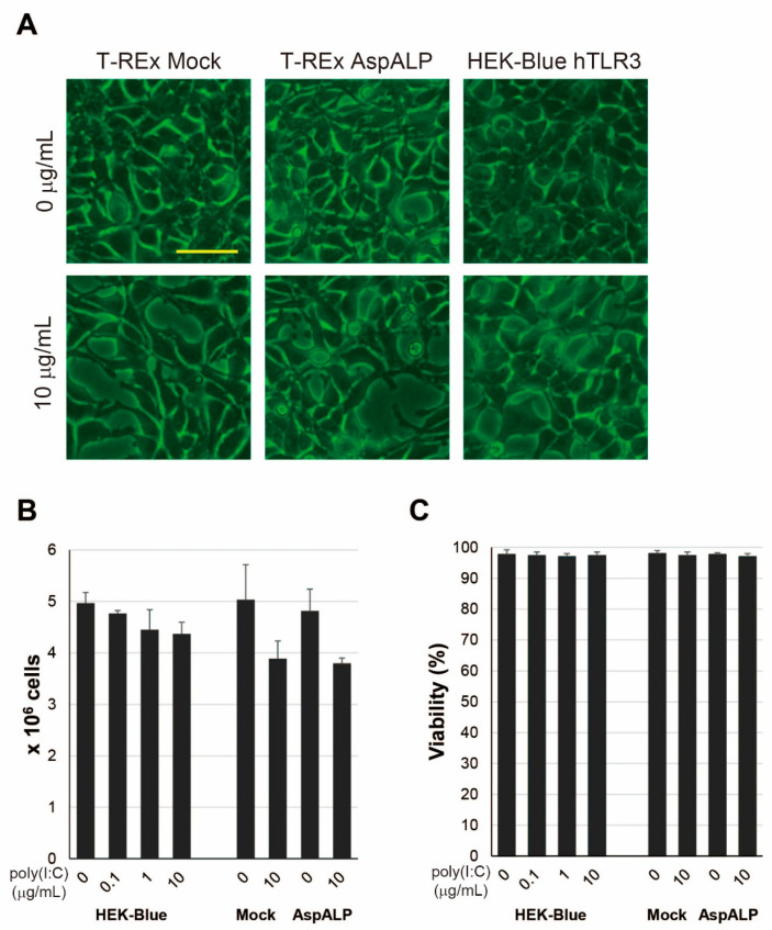
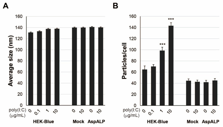
Signal peptides (SPs) not only mediate targeting to the endoplasmic reticulum (ER) but also play important roles as biomarkers and substances with physiological activity in extracellular fluids including blood. SPs are thought to be degraded intracellularly, making it unclear how they are transported from the ER to the extracellular fluid. In a recent study, we showed that a C-terminal fragment of the SP of a type I membrane protein, amyloid precursor protein (APP), was secreted into the extracellular fluid via exosomes using transformed HEK293 cells expressing APP SP flanking a reporter protein. In the present study, we demonstrate that a N-terminal fragment of the SP from a type II membrane protein, human placental secreted alkaline phosphatase (SEAP), is contained in exosomes and secreted into the extracellular fluid using HEK-Blue hTLR3 cells, which express both a human toll-like receptor 3 gene and an inducible SEAP reporter gene. When HEK-Blue hTLR3 cells were stimulated with a TLR3 ligand, a N-terminal fragment of SEAP SP in exosomes was increased in parallel with SEAP secretion in a concentration-dependent manner. These results indicated that SP fragments are exosomal components. In addition, migrating SP fragments were determined by characteristics of the signal-anchor sequence of membrane proteins. Furthermore, we found that SP fragments could bind to calmodulin (CALM), which is a cytosolic protein and also a component of exosomes, suggesting its involvement in the transportation of SP fragments from the endoplasmic reticulum to exosomes.

Keywords: calmodulin; exosomes; intercellular communication; signal peptide.


Conflict of interest statement

The authors declare no conflict of interest.

Figures

Figure 1
Figure 1
SEAP is released from poly(I:C)-stimulated cells. (A) The SEAP activity of T-REx Mock (Mock) and T-REx AspALP (AspALP) cells in the presence or absence of Dox at 1 μg/mL or poly(I:C) at 10 μg/mL. *** p < 0.001 vs. T-REx Mock cells in the absence of poly(I:C). ### p < 0.001 vs. T-REx AspALP cells in the absence of Dox. (B) The SEAP activity of HEK-Blue hTLR3 cells in the presence of poly(I:C) at 0–10 μg/mL. *** p < 0.001 vs. HEK-Blue hTLR3 cells in the absence of poly(I:C). (C) The peptide solution extracted from exosomes of HEK-Blue hTLR3 cells 3 days after poly(I:C) treatment at 10 μg/mL and T-REx AspALP cells 3 days after Dox treatment at 1 μg/mL was analyzed by MALDI-TOF MS.
Figure 2
Figure 2
N-terminal fragments of SEAP SP in exosomes released from HEK-Blue hTLR3 cells. (A) The peptide solution extracted from exosomes of HEK-Blue hTLR3 cells in the presence of poly(I:C) at 0–10 μg/mL for three days was analyzed by MALDI-TOF MS. (B) MS/MS analysis of peaks at m/z 1277 +/− 6 was performed. (C) The peak intensity at 1277 was measured by MALDI-TOF MS. ** p < 0.01, *** p < 0.001 vs. the absence of poly(I:C).
Figure 3
Figure 3
Morphology and growth in cells stimulated with poly(I:C). (A) Morphology of T-REx Mock (Mock), T-REx AspALP (AspALP), and HEK-Blue hTLR3 (HEK-Blue) cells after three days in the presence or absence of poly(I:C) at 10 μg/mL. Scale bar indicates 50 μm. The number (B) and viability (C) of cells after three days in the presence or absence of poly(I:C) at 0–10 μg/mL.
Figure 4
Figure 4
Properties of exosomes released from poly(I:C)-stimulated cells. (A) The average size of exosomes released from poly(I:C)-stimulated HEK-Blue hTLR3, T-REx Mock and T-REx AspALP cells. (B) Released exosomes per cell. *** p < 0.001 vs. cells in the absence of poly(I:C).
Figure 5
Figure 5
Exosomes do not contaminate the ER fraction from HEK-Blue hTLR3 cells. (A) Western blot analysis of markers for the ER and exosomes (Exo) fractions from HEK-Blue hTLR3 cells in the presence or absence of poly(I:C). (B) The ER fraction from HEK-Blue hTLR3 cells after poly(I:C) treatment was analyzed by MALDI-TOF MS.
Figure 6
Figure 6
Binding of exosomal SP fragments with CALM in the presence of Ca2+. (A) The peptide solution extracted from His-tagged CALM was analyzed by MALDI-TOF MS. (B) The peak intensity at 861 was measured by MALDI-TOF MS. ND, not detected. *** p < 0.001 vs. CALM(+)/APP SP(+)/Ca2+(−). (C) MS/MS analysis of the peaks at m/z 859 +/− 4. (D) The peak intensity at 861 and 2073 in the presence or absence of CALMIP was measured by MALDI-TOF MS. ND, not detected. *** p < 0.001 vs. CALM(+)/APP SP(+)/Ca2+(+)/CALMIP(−); (E) the working hypothesis of this study.

Similar articles

Cited by

References

    1. Pemberton C.J., Siriwardena M., Kleffmann T., Ruygrok P., Palmer S.C., Yandle T.G., Richards A.M. First identification of circulating prepro-A-type natriuretic peptide (preproANP) signal peptide fragments in humans: Initial assessment as cardiovascular biomarkers. Clin. Chem. 2012;58:757–767. doi: 10.1373/clinchem.2011.176990. - DOI - PubMed
    1. Siriwardena M., Kleffmann T., Ruygrok P., Cameron V.A., Yandle T.G., Nicholls M.G., Richards A.M., Pemberton C.J. B-type natriuretic peptide signal peptide circulates in human blood: Evaluation as a potential biomarker of cardiac ischemia. Circulation. 2010;122:255–264. doi: 10.1161/CIRCULATIONAHA.109.909937. - DOI - PubMed
    1. Pemberton C.J., Siriwardena M., Kleffmann T., Richards A.M. C-type natriuretic peptide (CNP) signal peptide fragments are present in the human circulation. Biochem. Biophys. Res. Commun. 2014;449:301–306. doi: 10.1016/j.bbrc.2014.05.020. - DOI - PubMed
    1. Wu C.M., Chang M.D.T. Signal peptide of eosinophil cationic protein is toxic to cells lacking signal peptide peptidase. Biochem. Biophys. Res. Commun. 2004;322:585–592. doi: 10.1016/j.bbrc.2004.07.160. - DOI - PubMed
    1. Chang H.T., Kao Y.L., Wu C.M., Fan T.C., Lai Y.K., Huang K.L., Chang Y.S., Tsai J.J., Chang M.D.T. Signal peptide of eosinophil cationic protein upregulates transforming growth factor-alpha expression in human cells. J. Cell. Biochem. 2007;100:1266–1275. doi: 10.1002/jcb.21120. - DOI - PubMed

Substances

LinkOut - more resources