. 2022 Mar 15;23(6):3163.
doi: 10.3390/ijms23063163.

cADPR Does Not Activate TRPM2

Affiliations

cADPR Does Not Activate TRPM2

Winnie Maria Riekehr et al. Int J Mol Sci. .

Abstract

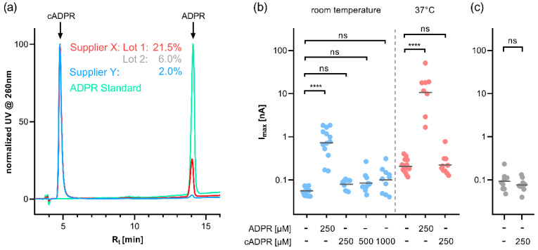
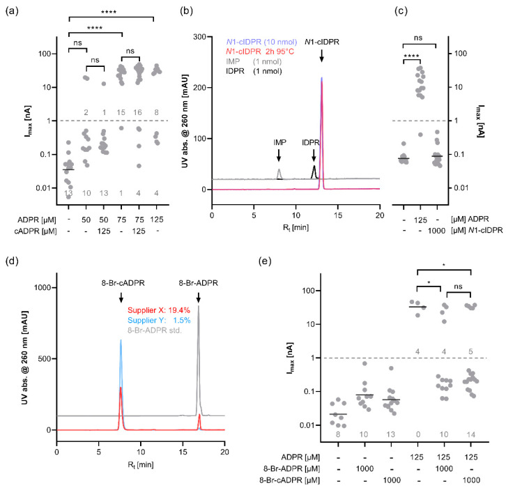
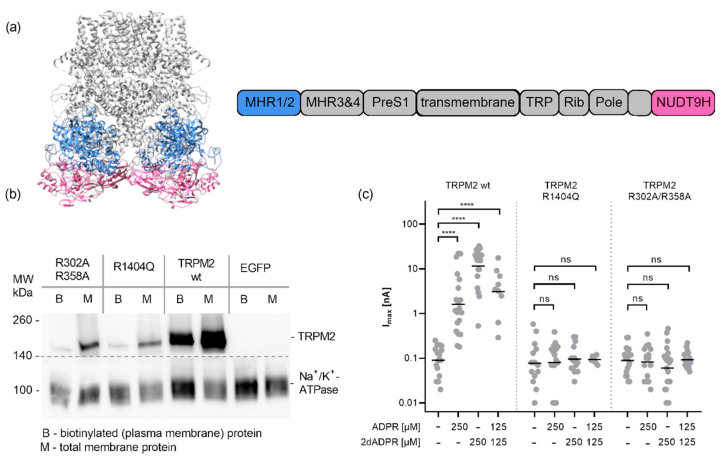
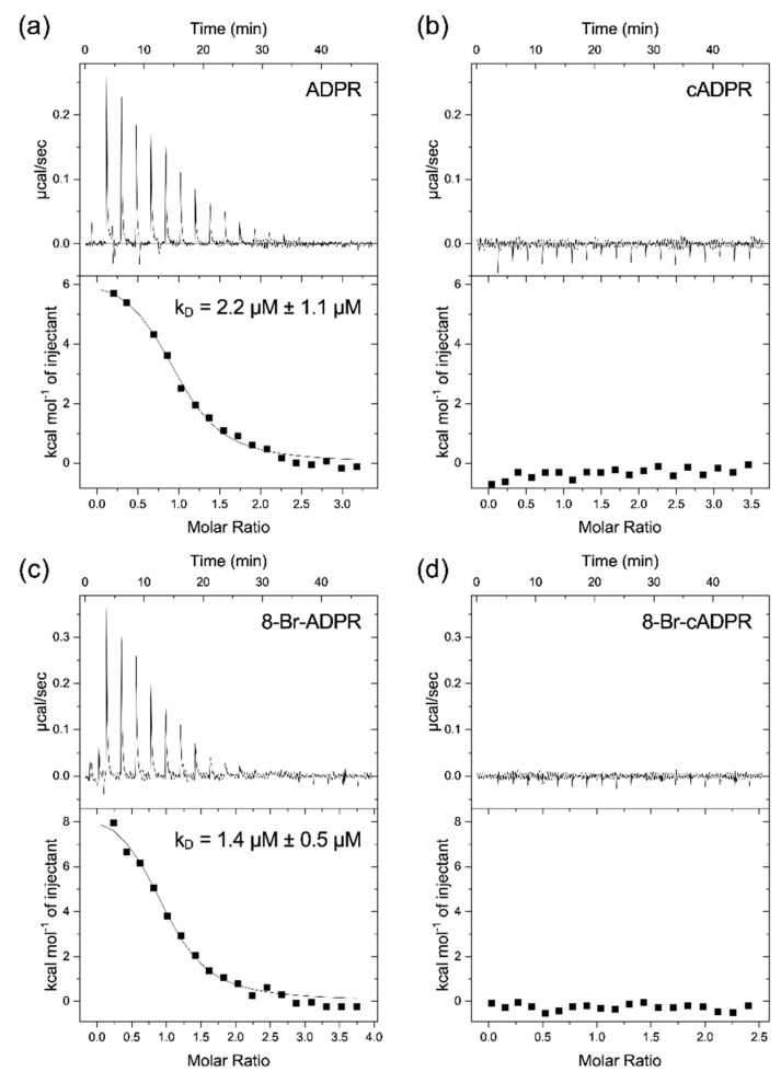
cADPR is a second messenger that releases Ca2+ from intracellular stores via the ryanodine receptor. Over more than 15 years, it has been controversially discussed whether cADPR also contributes to the activation of the nucleotide-gated cation channel TRPM2. While some groups have observed activation of TRPM2 by cADPR alone or in synergy with ADPR, sometimes only at 37 °C, others have argued that this is due to the contamination of cADPR by ADPR. The identification of a novel nucleotide-binding site in the N-terminus of TRPM2 that binds ADPR in a horseshoe-like conformation resembling cADPR as well as the cADPR antagonist 8-Br-cADPR, and another report that demonstrates activation of TRPM2 by binding of cADPR to the NUDT9H domain raised the question again and led us to revisit the topic. Here we show that (i) the N-terminal MHR1/2 domain and the C-terminal NUDT9H domain are required for activation of human TRPM2 by ADPR and 2'-deoxy-ADPR (2dADPR), (ii) that pure cADPR does not activate TRPM2 under a variety of conditions that have previously been shown to result in channel activation, (iii) the cADPR antagonist 8-Br-cADPR also inhibits activation of TRPM2 by ADPR, and (iv) cADPR does not bind to the MHR1/2 domain of TRPM2 while ADPR does.

Keywords: calcium signalling; cyclic adenosine 5′-diphosphate ribose; second messenger; transient receptor potential channel.


Conflict of interest statement

The authors declare no conflict of interest.

Figures

Figure 1
Figure 1
(a) HPLC analysis of cADPR from different suppliers (red, grey and blue). Due to the varying amount of cADPR in the preparations, the chromatograms have been normalised to the cADPR peak to illustrate the different fractions of ADPR in the preparations. The ADPR standard (green) is shown for comparison. (b) cADPR does not activate human TRPM2 at either room temperature or at 37 °C. HEK293 cells with stable expression of human TRPM2 were infused via the patch pipette with an intracellular solution buffered to a Ca2+ of 200 nM with EGTA and containing either 250 µM ADPR or 250 µM cADPR. Cells were kept at either room temperature (blue) or 37 °C (red) by continuous perfusion with bath solution. The bath solution contained NMDG instead of Na+. (See Supplementary Material Figure S1 for representative time courses and a schematic representation of the voltage ramp) (c) cADPR does not activate human TRPM2 in a bath solution with NaCl. At room temperature, TRPM2-expressing HEK293 cells were infused with an intracellular solution that contained either no nucleotide or 250 µM cADPR. Max currents at +15 mV from repetitive voltage ramps are shown on a log scale. Log transformed data were tested against the respective control by one-way ANOVA followed by post hoc t test with Bonferroni correction (b) or two-tailed Student’s t test (c), the mean of the log-transformed data is indicated by a horizontal bar (**** p < 0.0001, ns not significant).
Figure 2
Figure 2
(a) cADPR does not enhance the TRPM2 current at threshold concentrations of ADPR. HEK293 cells with stable expression of human TRPM2 were infused via the patch pipette with an intracellular solution with weak Ca2+ buffering (100 µM EGTA) and containing varying concentrations of ADPR with or without 125 µM cADPR. During the recording, cells were kept at room temperature. With ADPR in the pipette solution, the currents show a bimodal distribution, with some cells showing a slight increase in current compared with the control while others exhibit a current two orders of magnitude higher. Log-transformed data were tested using Kruskal–Wallis test with Dunn’s correction (**** adj. p < 0.0001, ns = not significant) (b) HPLC analysis of N1-cIDPR (blue), N1-cIDPR that has been incubated at 95 °C for 2 h (red), and standards of potential breakdown products IMP (grey) and IDPR (black). (c) The nonhydrolysable cADPR analogue N1-cIDPR does not activate human TRPM2. TRPM2-expressing HEK293 cells were infused with a pipette solution containing either no nucleotide or either ADPR or N1-cIDPR. Log-transformed data were tested using Kruskal–Wallis test with Dunn’s correction (**** adj. p <0.0001, ns = not significant) (d) HPLC analysis of 8-Br-cADPR from two different suppliers (red and blue) and an 8-Br-ADPR standard (grey). The numbers indicate the fractional peak area of the 8-Br-ADPR peak in the analysed 8-Br-cADPR. (e) 8-Br-ADPR and 8-Br-cADPR reduce the ADPR-induced current in TRPM2-expressing HEK293 cells. Cells were infused with a pipette solution that contained no nucleotide, 125 µM ADPR, 1 mM 8-Br-ADPR, 1 mM 8-Br-cADPR, or a combination thereof. For some conditions, the horizontal bar indicates the mean of the log-transformed currents. Due to the bimodal distribution, the fraction of cells responding to a current above and below 1 nA were compared using Fisher’s exact test (* p < 0.05, ns = not significant).
Figure 3
Figure 3
Effect of mutations in the MHR1/2 domain and in the NUDT9H domain on activation of TRPM2 by ADPR and 2′-deoxy-ADPR. (a) Structure of TRPM2, visualization of PDB:6PUS [18] generated in UCSF chimera [34], MHR1/2 (blue) and NUDT9H (magenta) domains are highlighted. (b) Effect of mutations on the plasma membrane localisation of TRPM2. Transiently transfected HEK293 cells (as above, additionally, empty pIRES-EGFP vector control was used) were treated with a nonmembrane permeant biotinylating agent. Cells were harvested and total membrane proteins isolated (M). The biotinylated proteins were enriched using neutravidin agarose beads. After separation on a 4–15% SDS-PAGE proteins were transferred to a PVDF membrane and detected by chemoluminescence using primary antibodies against human TRPM2 (upper part, NB 500-241, Novus Biologicals) and human Na+/K+-ATPase ((lower part, #3010, Cell Signalling Technology) and an HRP-conjugated secondary antibody, both parts, Dianova #111-035-045) (c) HEK293 cells were either transiently transfected with expression vectors for human TRPM2 with mutations in either the NUDT9H domain (R1404Q) or in the MHR1/2 domain (R30/R358A) or the wild type channel. 24 h post-transfection cells were placed in a bath solution with NMDG and infused via the patch pipette with an intracellular solution buffered to a Ca2+ of 200 nM with EGTA and containing either ADPR, 2′-deoxy-ADPR or a combination of both. Max currents at +15 mV obtained from repetitive voltage ramps are shown on a log scale. Log-transformed data were tested against the respective control by one-way ANOVA followed by a post hoc t test with Bonferroni correction; the mean is indicated by a horizontal bar (**** p < 0.0001, ns not significant).
Figure 4
Figure 4
Binding of ADPR, cADPR, 8-Br-ADPR, and 8-Br-cADPR to the isolated MHR1/2 domain from drTRPM2. The isolated MHR1/2 domain from drTRPM2 was expressed in E. coli and purified by affinity and size exclusion chromatography and used to determine the binding affinity by isothermal titration calorimetry. The figure shows representative raw data and integrated peaks from 3 experiments per condition (a) ADPR, (b) cADPR, (c) 8-Br-ADPR, and (d) 8-Br-cADPR. The KD value is expressed as mean ± SEM (n = 3). Raw data from individual experiments can be found in Supplementary Material Figure S2.

References

    1. Clapper D.L., Walseth T.F., Dargie P.J., Lee H.C. Pyridine nucleotide metabolites stimulate calcium release from sea urchin egg microsomes desensitized to inositol trisphosphate. J. Biol. Chem. 1987;262:9561–9568. doi: 10.1016/S0021-9258(18)47970-7. - DOI - PubMed
    1. Lee H.C. Physiological Functions of Cyclic ADP-Ribose and NAADP as Calcium Messengers. Annu. Rev. Pharmacol. Toxicol. 2001;41:317–345. doi: 10.1146/annurev.pharmtox.41.1.317. - DOI - PubMed
    1. Lee H.C. The Cyclic ADP-Ribose/NAADP/CD38-Signaling Pathway: Past and Present. Messenger. 2012;1:16–33. doi: 10.1166/msr.2012.1005. - DOI
    1. Galione A., Lee H.C., Busa W.B. Ca(2+)-induced Ca2+ release in sea urchin egg homogenates: Modulation by cyclic ADP-ribose. Science. 1991;253:1143–1146. doi: 10.1126/science.1909457. - DOI - PubMed
    1. Pérez C.F., Marengo J.J., Bull R., Hidalgo C. Cyclic ADP-ribose activates caffeine-sensitive calcium channels from sea urchin egg microsomes. Am. J. Physiol. 1998;274:C430–C439. doi: 10.1152/ajpcell.1998.274.2.C430. - DOI - PubMed