

Adipose-Derived Stem Cells Preincubated with Green Tea EGCG Enhance Pancreatic Tissue Regeneration in Rats with Type 1 Diabetes through ROS/Sirt1 Signaling Regulation
- PMID: 35328586
- PMCID: PMC8951845
- DOI: 10.3390/ijms23063165
Adipose-Derived Stem Cells Preincubated with Green Tea EGCG Enhance Pancreatic Tissue Regeneration in Rats with Type 1 Diabetes through ROS/Sirt1 Signaling Regulation
Abstract
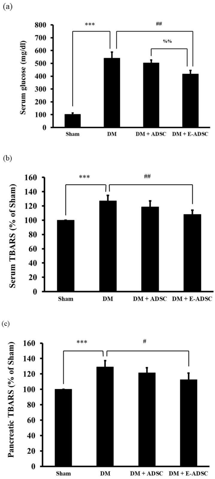
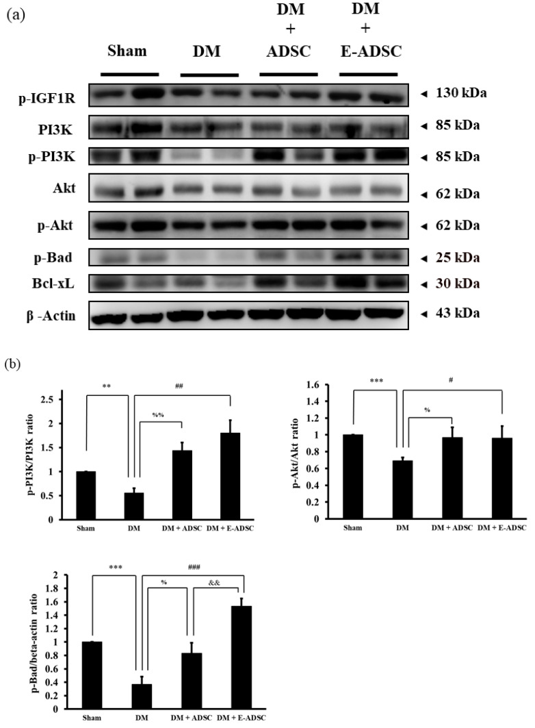
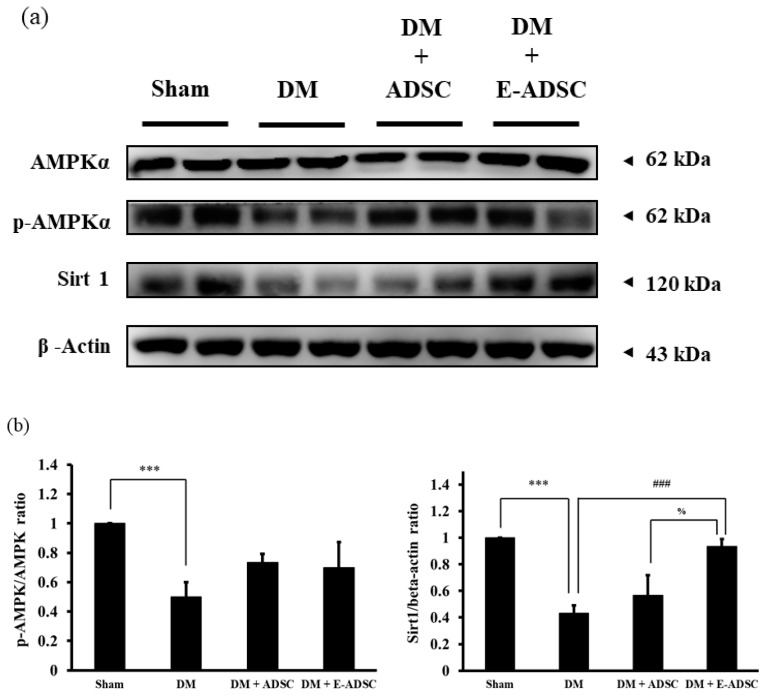
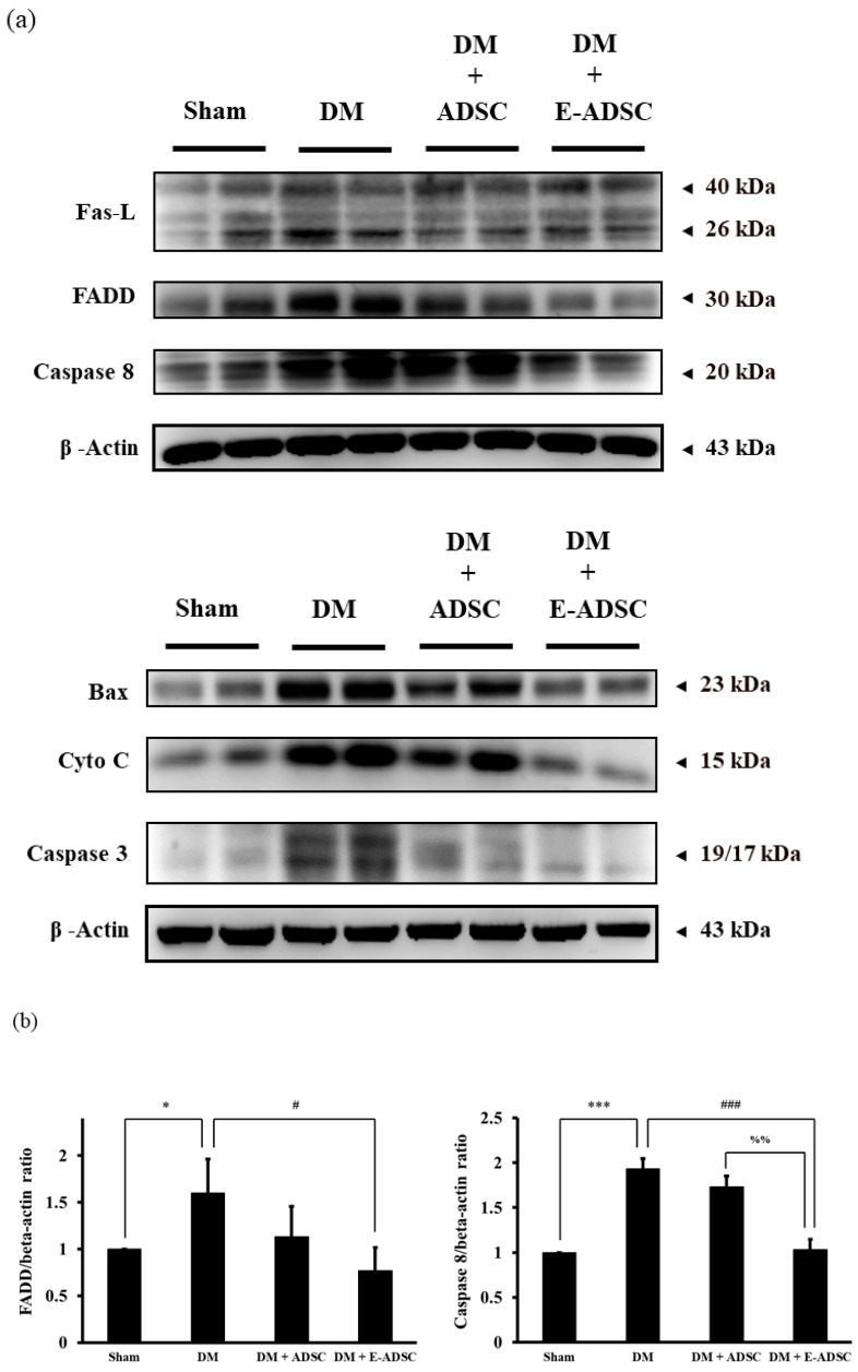
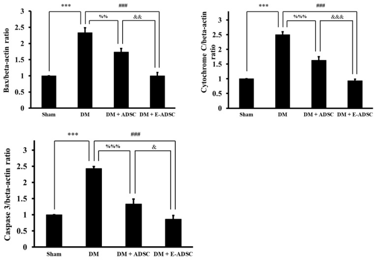
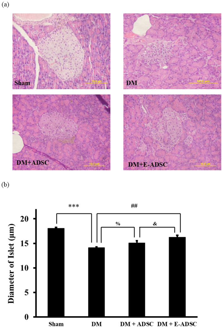
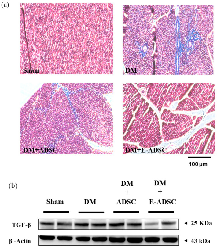
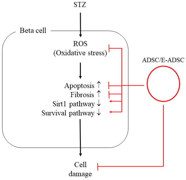
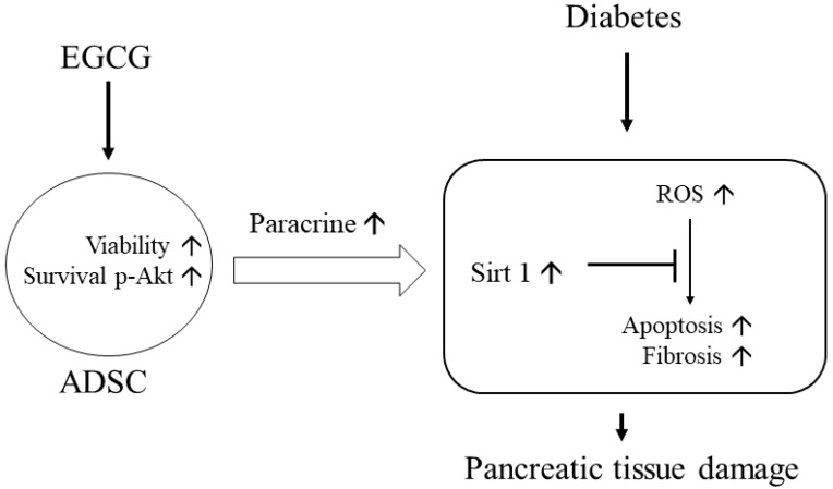
Type 1 diabetes stem-cell-based therapy is one of the best therapeutic approaches for pancreatic damage treatment due to stem cell tissue regeneration. Epigallocatechin gallate (EGCG) is one of the active components found in green tea. Experimental results suggest that EGCG shows beneficial effects on cell protection. This study explores whether a better pancreatic regeneration therapeutic effect could be found in mesenchymal stem cells pretreated with EGCG compared to stem cells without EGCG pretreatment. A cell model confirmed that adipose-derived stem cells (ADSC) incubated with EGCG increase cell viability under high-glucose (HG) stress. This is due to survival marker p-Akt expression. In an animal model, type 1 diabetes induced the activation of several pathological signals, including islet size reduction, extracellular fibrotic collagen deposition, oxidative stress elevation, survival pathway suppression, apoptosis signaling induction, and Sirt1 antioxidant pathway downregulation. Ordinary ADSC transplantation slightly improved the above pathological signals. Further, EGCG-pretreated ADSC transplantation significantly improved the above pathological conditions. Taken together, EGCG-pretreated ADSCs show clinical potential in the treatment of patients with type 1 diabetes through the regeneration of damaged pancreatic tissues.
Keywords: green tea EGCG; mesenchymal stem cells; pancreatic regeneration; survival p-Akt.
Conflict of interest statement
The authors declare no conflict of interest.
Figures
Similar articles
-
Autologous transplantation of green tea epigallocatechin-3-gallate pretreated adipose-derived stem cells increases cardiac regenerative capability through C-X-C motif chemokine receptor 4 expression in the treatment of rats with diabetic cardiomyopathy.Exp Anim. 2024 Jul 9;73(3):246-258. doi: 10.1538/expanim.23-0109. Epub 2024 Mar 7. Exp Anim. 2024. PMID: 38447976 Free PMC article.
-
Oral administration of green tea Epigallocatechin-3-gallate reduces oxidative stress and enhances restoration of cardiac function in diabetic rats receiving autologous transplantation of adipose-derived stem cells.Arch Physiol Biochem. 2021 Feb;127(1):82-89. doi: 10.1080/13813455.2019.1614631. Epub 2019 May 21. Arch Physiol Biochem. 2021. PMID: 31112046
-
Green tea epigallocatechin gallate enhances cardiac function restoration through survival signaling expression in diabetes mellitus rats with autologous adipose tissue-derived stem cells.J Appl Physiol (1985). 2017 Nov 1;123(5):1081-1091. doi: 10.1152/japplphysiol.00471.2016. Epub 2017 May 25. J Appl Physiol (1985). 2017. PMID: 28546469
-
Epigallocatechin-Gallate: Unraveling Its Protective Mechanisms and Therapeutic Potential.Cell Biochem Funct. 2025 Feb;43(2):e70056. doi: 10.1002/cbf.70056. Cell Biochem Funct. 2025. PMID: 39915982 Review.
-
Polyphenolic antioxidant (-)-epigallocatechin-3-gallate from green tea reduces UVB-induced inflammatory responses and infiltration of leukocytes in human skin.Photochem Photobiol. 1999 Feb;69(2):148-53. Photochem Photobiol. 1999. PMID: 10048310 Review.
Cited by
-
Ohwia caudata aqueous extract attenuates senescence in aging adipose-derived mesenchymal stem cells.Heliyon. 2024 Apr 16;10(9):e29729. doi: 10.1016/j.heliyon.2024.e29729. eCollection 2024 May 15. Heliyon. 2024. PMID: 38698985 Free PMC article.
-
The Role of Catechins in Regulating Diabetes: An Update Review.Nutrients. 2022 Nov 4;14(21):4681. doi: 10.3390/nu14214681. Nutrients. 2022. PMID: 36364943 Free PMC article. Review.
-
Trifolium pratense-Derived Exosome Improved Serum Biochemical Parameters and Pancreatic Genes in STZ-Induced Diabetic Rats.Endocrinol Diabetes Metab. 2025 Sep;8(5):e70103. doi: 10.1002/edm2.70103. Endocrinol Diabetes Metab. 2025. PMID: 40887321 Free PMC article.
-
L-Theanine-Treated Adipose-Derived Mesenchymal Stem Cells Alleviate the Cytotoxicity Induced by N-Nitrosodiethylamine in Liver.Tissue Eng Regen Med. 2022 Dec;19(6):1207-1221. doi: 10.1007/s13770-022-00472-2. Epub 2022 Aug 27. Tissue Eng Regen Med. 2022. PMID: 36029414 Free PMC article.
-
Natural small molecules synergize mesenchymal stem cells for injury repair in vital organs: a comprehensive review.Stem Cell Res Ther. 2024 Aug 7;15(1):243. doi: 10.1186/s13287-024-03856-4. Stem Cell Res Ther. 2024. PMID: 39113141 Free PMC article. Review.
References
MeSH terms
Substances
LinkOut - more resources
Full Text Sources
Medical
