. 2022 Mar 15;23(6):3178.
doi: 10.3390/ijms23063178.

CRISPR/Cas9-Based Mutagenesis of Histone H3.1 in Spinal Dynorphinergic Neurons Attenuates Thermal Sensitivity in Mice

Affiliations

CRISPR/Cas9-Based Mutagenesis of Histone H3.1 in Spinal Dynorphinergic Neurons Attenuates Thermal Sensitivity in Mice

Zoltán Mészár et al. Int J Mol Sci. .

Abstract

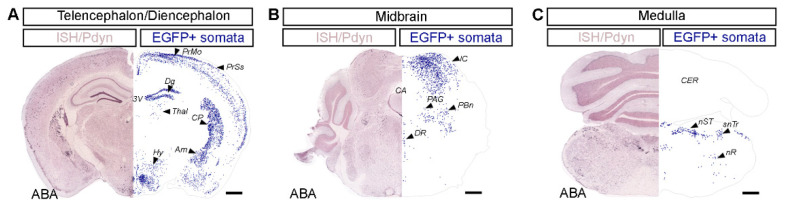
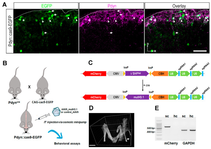
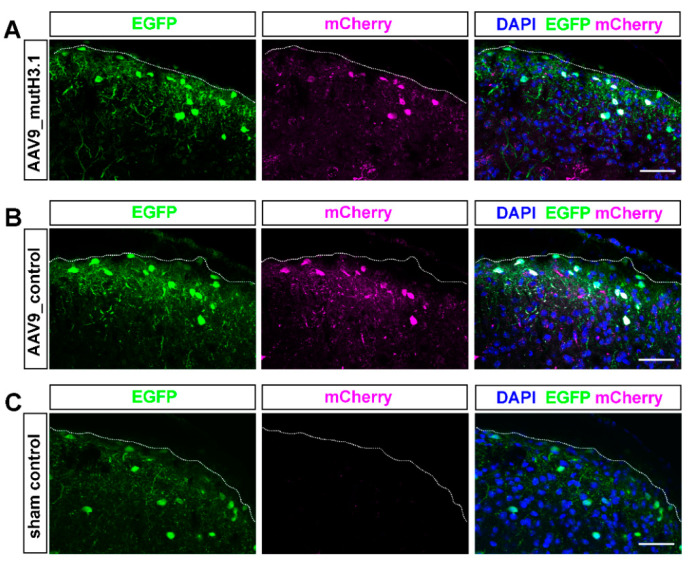
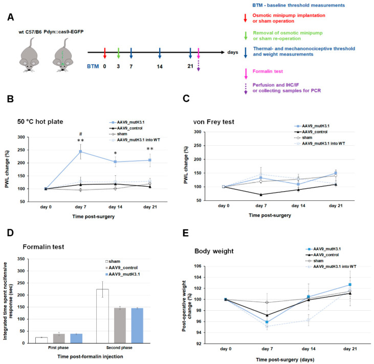
Burn injury is a trauma resulting in tissue degradation and severe pain, which is processed first by neuronal circuits in the spinal dorsal horn. We have recently shown that in mice, excitatory dynorphinergic (Pdyn) neurons play a pivotal role in the response to burn-injury-associated tissue damage via histone H3.1 phosphorylation-dependent signaling. As Pdyn neurons were mostly associated with mechanical allodynia, their involvement in thermonociception had to be further elucidated. Using a custom-made AAV9_mutH3.1 virus combined with the CRISPR/cas9 system, here we provide evidence that blocking histone H3.1 phosphorylation at position serine 10 (S10) in spinal Pdyn neurons significantly increases the thermal nociceptive threshold in mice. In contrast, neither mechanosensation nor acute chemonociception was affected by the transgenic manipulation of histone H3.1. These results suggest that blocking rapid epigenetic tagging of S10H3 in spinal Pdyn neurons alters acute thermosensation and thus explains the involvement of Pdyn cells in the immediate response to burn-injury-associated tissue damage.

Keywords: dynorphinergic neuron; epigenetic regulation; heat sensation; histone; pain; spinal cord.


Conflict of interest statement

The authors have declared that no conflict of interest exists.

Figures

Figure 1
Figure 1
Immunohistochemical mapping revealed that regional distribution of dynorphinergic neurons in the brain of a Pdyn::cas9-EGFP mouse shows large similarity to the reference atlas provided by the Allen Institute. The distribution of Pdyn neurons was evaluated by plotting immunopositive cell bodies (revealed by the HRP/DAB method) through the telencephalon/diencephalon (A), the midbrain (B), and the medulla (C) of a Pdyn::cas9-EGFP mouse. On the right side of each panel, blue dots represent the location of Pdyn-immunopositive neurons, detected and reconstructed with the aid of Neurolucida (EGFP + somata). The left side of each panel shows the corresponding reference image of in situ hybridization (ISH) data from the Allen Brain Atlas (ABA) [22] displaying Pdyn mRNA expression pattern. PrMo, primary motor area; PrSs, primary somatosensory area layer 2/3 and layer 5; Dg, dentate gyrus; Thal, mediodorsal nucleus of the thalamus; Hy, dorsomedial nucleus of the hypothalamus; CP, caudoputamen; Am, central amygdalar nucleus; 3V, third ventricle; IC, inferior colliculus; PAG, periaqueductal gray; PBn, parabrachial nucleus; DR, dorsal nucleus raphe; CA, cerebral aqueduct; CER, cerebellum; nST, nucleus of the solitary tract; snTr, spinal nucleus of the trigeminal; nR, reticular nucleus. Scale bar, 500 µm. For original images, see Supplementary Figure S1.
Figure 2
Figure 2
Cell-type specific targeted delivery of the necessary components for genome editing of histone H3.1 via CRISPR/cas9 strategy. (A) Immunostaining with antibodies against EGRP (green), Pdyn (magenta) in a projected image of seven optical sections with a 40× lens from a transverse spinal cord section of a Pdyn::cas9-EGFP mouse. The overlay shows a merged image. The numerous EGRP-immunoreactive neurons are predominantly visible in the superficial layers of the lumbar spinal cord. The majority of these are Pdyn+ (arrowheads), while some of them lack Pdyn (arrow). Few Pdyn-expressing neurons lack the EGFP signal (asterisk). D, dorsal; M, medial; scale bar, 50 μm. (B) Schematic drawing representing the CRISPR/cas9 strategy to establish the mutant histone H3.1 (mutH3.1) in Pdyn neurons. The abbreviation mutH3.1 refers to serine-to-alanine exchange (S10A) at position serine 10 of the wild-type histone H3.1. IT, intrathecal; AAV9_mutH3.1, the mutant histone H3.1-containing recombinant adenoassociated virus serotype 9. (C) Schematic representation of the final insert synthesized and cloned into a recombinant AAV9. This cassette encoded mutH3.1 flanked by loxP sites (purple), three single-guide RNAs (sgRNAs; blue) driven by the human polymerase III U6 promoters (green), and a mCherry fluorescent protein (red). In this approach, S10A point mutation would be introduced into only cre-expressing neurons (i.e., into Pdyn-expressing neurons in Pdyn::cas9-EGFP hybrids). CMV, human cytomegalovirus (CMV) immediate early enhancer and promoter; CBH, chicken beta-actin promoter with CMV enhancer. For sgRNA sequences targeting wild-type histone H3.1, see Supplementary Table S3. See also Supplementary Data S1 for further technical details. (D) 3D volume reconstruction of micro-CT images, used for validating the intrathecal position of the inserted cannula before the osmotic pump implantation. The intrathecal catheter (arrow) is shown within the subarachnoid space in a living deeply anesthetized Pdyn::cas9-EGFP mouse. The catheter was introduced at the level of L5-L6 vertebral laminae and pushed up to L1-L2. Scale bar, 5000 μm. (E) In contrast to the hippocampus (hc), mCherry-specific RT-PCR produced a single sharp band in the spinal cord (sc) sample of a wild-type mouse that had been transfected with the AAV9_mutH3.1. GAPDH was amplified in both samples. BenchTop 100 bp DNA ladder was used as a reference.
Figure 3
Figure 3
Distribution of AAV9 viral particle in the spinal dorsal horn of Pdyn::cas9-EGFP mice as identified by the presence of mCherry tag encoded by the virus. Representative images showing immunostaining with antibodies against EGFP (green), mCherry (magenta), and DAPI (blue) in projected images of 10 optical sections (each 0.5-µm-thick) taken with a 40× lens in the spinal cord from an AAV9_mutH3.1 vector treated (A), an AAV9_control treated (B) and a sham-operated (C) Pdyn::cas9-EGFP mouse. (A) Neurons showing mCherry-immunoreactivity are scattered throughout the SDH. Some Pdyn neurons (green to to their EGFP expression) show strong mCherry signal especially in the superficial region of the dorsal horn. (B) Administration of AAV9_control virus into Pdyn::cas9-EGFP mice produced an expression pattern of mCherry, similar to that shown in panel A. (C) The mCherry-specific fluorescent signal is completely missing in the transverse spinal cord sections of animals in the sham-operated group. White dotted lines indicate borders between white and gray matter. Scale bars is 50 µm.
Figure 4
Figure 4
Intrathecal administration of AAV9_mutH3.1 into Pdyn::cas9-EGFP mice increases the thermal nociceptive threshold. (A) Schematic time scale of the experimental procedures. WT, wild-type C57Bl/6; IHC, immunohistochemistry; IF, immunofluorescent staining. (B,C) Changes in paw withdrawal latencies (PWL) to thermal- and mechanical pain were evaluated before (day 0) and after the surgery (day 7, 14, 21) in different groups of Pdyn::cas9-EGFP mice (i.e., AAV9_mutH3.1, AAV9_control, sham-operated) and in wild-type animals transduced with the AAV9_mutH3.1. Values at Day 0 represent the pre-surgery baseline values (BTM). (B) AAV9_mutH3.1-treated animals exhibited higher thermal nociceptive threshold compared to the AAV9_control and sham operated groups at day 7. This significant elevation was persistent until the end of the observational period. * p < 0.05 and ** p < 0.01 compared with the AAV9_control group (details in Supplementary Table S4). # p < 0.01 when the overall influence of the treatment with the AAV9_mutH3.1 on paw withdrawal latency (PWL) in response to noxious heat compared with the pre-surgery baseline (p = 0.009, n = 7, Kruskal–Wallis ANOVA). (C) Paw withdrawal latency to painful mechanical stimuli showed no significant alterations within, and differences between the groups. (D) Formalin-induced somatic pain was quantified as the integrated time spent exhibiting nocifensive behavioral during early (0–15 min) and late (15–60 min) phases of formalin application. Formalin-induced nocifensive behavior was reduced by 40% in the second phase in mice that had been infected with AAV9 irrespectively from their transgenes to be expressed, although, this reduction did not reach a statistically significant level. For additional details for statistical comparison see Supplementary Table S4. See also Supplementary Figure S5 for raw data of the formalin-induced nocifensive behavior. (E) Changes in body weight were evaluated before (day 0) and after the surgery (day 7, 14, 21) in different groups of Pdyn::cas9-EGFP mice (i.e., AAV9_mutH3.1, AAV9_control, sham-operated). Day 0 represents the pre-surgery baseline value. Transduction with the viruses (AAV9_mutH3.1 or AAV9_control) led to a modest body weight loss by day 7 that resolved later in all groups.

Similar articles

Cited by

References

    1. Liang D.Y., Sun Y., Shi X.Y., Sahbaie P., Clark J.D. Epigenetic regulation of spinal cord gene expression controls opioid-induced hyperalgesia. Mol. Pain. 2014;10:59. doi: 10.1186/1744-8069-10-59. - DOI - PMC - PubMed
    1. Descalzi G., Ikegami D., Ushijima T., Nestler E.J., Zachariou V., Narita M. Epigenetic mechanisms of chronic pain. Trends Neurosci. 2015;38:237–246. doi: 10.1016/j.tins.2015.02.001. - DOI - PMC - PubMed
    1. Liang L., Tao Y.X. Expression of acetyl-histone H3 and acetyl-histone H4 in dorsal root ganglion and spinal dorsal horn in rat chronic pain models. Life Sci. 2018;211:182–188. doi: 10.1016/j.lfs.2018.09.029. - DOI - PubMed
    1. Tochiki K.K., Maiaru M., Norris C., Hunt S.P., Geranton S.M. The mitogen and stress-activated protein kinase 1 regulates the rapid epigenetic tagging of dorsal horn neurons and nocifensive behaviour. Pain. 2016;157:2594–2604. doi: 10.1097/j.pain.0000000000000679. - DOI - PMC - PubMed
    1. Torres-Perez J.V., Sántha P., Varga A., Szucs P., Sousa-Valente J., Gaal B., Nagy I. Phosphorylated Histone 3 at Serine 10 Identifies Activated Spinal Neurons and Contributes to the Development of Tissue Injury-Associated Pain. Sci. Rep. 2017;7:41221. doi: 10.1038/srep41221. - DOI - PMC - PubMed