. 2022 Mar 16;23(6):3213.
doi: 10.3390/ijms23063213.

4-Arylthiosemicarbazide Derivatives as Toxoplasmic Aromatic Amino Acid Hydroxylase Inhibitors and Anti-inflammatory Agents

Affiliations

4-Arylthiosemicarbazide Derivatives as Toxoplasmic Aromatic Amino Acid Hydroxylase Inhibitors and Anti-inflammatory Agents

Adrian Bekier et al. Int J Mol Sci. .

Abstract

Approximately one-third of the human population is infected with the intracellular cosmopolitan protozoan Toxoplasma gondii (Tg), and a specific treatment for this parasite is still needed. Additionally, the increasing resistance of Tg to drugs has become a challenge for numerous research centers. The high selectivity of a compound toward the protozoan, along with low cytotoxicity toward the host cells, form the basis for further research, which aims at determining the molecular targets of the active compounds. Thiosemicarbazide derivatives are biologically active organic compounds. Previous studies on the initial preselection of 58 new 4-arylthiosemicarbazide derivatives in terms of their anti-Tg activity and selectivity made it possible to select two promising derivatives for further research. One of the important amino acids involved in the proliferation of Tg and the formation of parasitophorous vacuoles is tyrosine, which is converted by two unique aromatic amino acid hydroxylases to levodopa. Enzymatic studies with two derivatives (R: para-nitro and meta-iodo) and recombinant aromatic amino acid hydroxylase (AAHs) obtained in the E. coli expression system were performed, and the results indicated that toxoplasmic AAHs are a molecular target for 4-arylthiosemicarbazide derivatives. Moreover, the drug affinity responsive target stability assay also confirmed that the selected compounds bind to AAHs. Additionally, the anti-inflammatory activity of these derivatives was tested using THP1-Blue™ NF-κB reporter cells due to the similarity of the thiosemicarbazide scaffold to thiosemicarbazone, both of which are known NF-κB pathway inhibitors.

Keywords: NF-κB pathway inhibition; Toxoplasma gondii; bradyzoite; thiosemicarbazide; tyrosine.


Conflict of interest statement

The authors declare no conflict of interest.

Figures
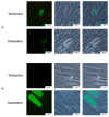
Figure 1
Figure 1
Confirmation of the differentiation of tachyzoites to bradyzoites by an immunofluorescent assay. (A) SAG1 tachyzoite-specific marker expression in Toxoplasma gondii ME49 strain. (B) MAG1 bradyzoite-specific marker expression in Toxoplasma gondii ME49 strain.
Figure 2
Figure 2
T. gondii aromatic amino acid hydroxylases expressed in this study. SDS–PAGE and Western blots of proteins expressed from AAH1 and AAH2. (A) SDS–PAGE of AAH1 and AAH2. (B) Monoclonal antibody anti-6xHis Tag. (C) Polyclonal antibody anti-TH. Due to the high degree of similarity with TH, recombinant toxoplasmic proteins are recognized by polyclonal antibodies against TH.
Figure 3
Figure 3
Lineweaver–Burk linearized plots with varying concentrations of compounds 2a and 6a. The concentration of inhibitor varied from 0 to 10 μM. As a substrate, L-tyrosine (L-Tyr) was used with 0.125, 0.25, 0.5, 1, and 2 mM concentrations. V—reaction rate. (A) AAH1 and 2a as inhibitors. (B) AAH2 and 2a as inhibitors. (C) AAH1 and 6a as inhibitors. (D) AAH2 and 6a as inhibitors. Each point represents the combined averages of three independent experiments in triplicate under the same conditions.
Figure 3
Figure 3
Lineweaver–Burk linearized plots with varying concentrations of compounds 2a and 6a. The concentration of inhibitor varied from 0 to 10 μM. As a substrate, L-tyrosine (L-Tyr) was used with 0.125, 0.25, 0.5, 1, and 2 mM concentrations. V—reaction rate. (A) AAH1 and 2a as inhibitors. (B) AAH2 and 2a as inhibitors. (C) AAH1 and 6a as inhibitors. (D) AAH2 and 6a as inhibitors. Each point represents the combined averages of three independent experiments in triplicate under the same conditions.
Figure 4
Figure 4
Protection of AAH1 from proteolysis by 4-arylthiosemicarbazide derivatives in the presence of 10 µM compound and with the Pronase:protein ratio. Western blots of AAH1 from the drug affinity responsive target stability assay and relative band intensity analysis. (A) Protection of AAH1 from proteolysis by 2a in the presence of 10 µM compound. (B) Protection of AAH1 from proteolysis by 6a in the presence of 10 µM compound. (C) Proteolytic activity inhibition by 2a protecting AAH1. (D) Proteolytic activity inhibition by 6a protecting AAH1. The intensities of the AAH1 bands were quantified using ImageJ software. Values with statistically significant differences are labeled by asterisks; **** p < 0.0001. Data were compared using one-way ANOVA with Tukey’s multiple comparisons test. Data were obtained from three independent experiments and are expressed as means ± standard deviations.
Figure 5
Figure 5
Illustration of the amount of stabilized AAH1 accessible for detection in the presence of increasing concentrations of 4-arylthiosemicarbazide derivatives. (A) The stabilization effect of 2a on AAH1 was evaluated by Western blot. (B) The stabilization effect of 6a on AAH1 was evaluated by Western blot. (C) Dose-dependence curve of 2a on AAH1. (D) Dose-dependence curve of 6a on AAH1. The intensities of the AAH1 bands were quantified using ImageJ software. The line was fitted with a three-parameter logistic curve. Data were obtained from three independent experiments and are expressed as the means ± SD.
Figure 5
Figure 5
Illustration of the amount of stabilized AAH1 accessible for detection in the presence of increasing concentrations of 4-arylthiosemicarbazide derivatives. (A) The stabilization effect of 2a on AAH1 was evaluated by Western blot. (B) The stabilization effect of 6a on AAH1 was evaluated by Western blot. (C) Dose-dependence curve of 2a on AAH1. (D) Dose-dependence curve of 6a on AAH1. The intensities of the AAH1 bands were quantified using ImageJ software. The line was fitted with a three-parameter logistic curve. Data were obtained from three independent experiments and are expressed as the means ± SD.
Figure 6
Figure 6
NF-κB induction in THP1-Blue™ human monocytes exposed to TLA, BLA, AAH1, or AAH2, or treated with LPS (positive control). Data are shown as the means from three repeats with error bars that indicate the standard deviations. Data were compared using a two-way ANOVA test, and for significant comparisons, further analysis was performed using Dunnett’s multiple comparisons test. Values with statistically significant differences are labeled by asterisks; **** means p < 0.0001. The differences were considered significant with a p value < 0.05 and are presented in Supplementary Materials Table S1. Aberration: nt—not treated; a.s. after stimulation; b.s. before stimulation; TLA—soluble tachyzoite antigens; BLA—soluble bradyzoite antigens; AAH—aromatic amino acid hydroxylases.
Figure 7
Figure 7
INF-γ production from supernatants after human monocytes were exposed to TLA, BLA, AAH1, or AAH2, or treated with LPS (positive control). Data are shown as the means from three repeats with error bars that indicate the standard deviations. Data were compared using a two-way ANOVA test, and for significant comparisons, further analysis was performed using Dunnett’s multiple comparisons test. Values with statistically significant differences are labeled by asterisks; **** p < 0.0001. The differences were considered significant when p values were less than 0.05, and such differences are presented in Supplementary Materials Table S1. Aberration: nt—not treated; a.s. after stimulation; b.s. before stimulation; TLA–soluble tachyzoite antigens; BLA—soluble bradyzoite antigens; AAH—aromatic amino acid hydroxylases.

Similar articles

Cited by

References

    1. Montoya J.G., Liesenfeld O. Toxoplasmosis. Lancet. 2004;363:1965–1976. doi: 10.1016/S0140-6736(04)16412-X. - DOI - PubMed
    1. Djurković-Djaković O., Dupouy-Camet J., Van der Giessen J., Dubey J.P. Toxoplasmosis: Overview from a One Health perspective. Food Waterborne Parasitol. 2019;15:e00054. doi: 10.1016/j.fawpar.2019.e00054. - DOI - PMC - PubMed
    1. Checkley W., White A.C., Jaganath D., Arrowood M.J., Chalmers R.M., Chen X.M., Fayer R., Griffiths J.K., Guerrant R.L., Hedstrom L., et al. A review of the global burden, novel diagnostics, therapeutics, and vaccine targets for cryptosporidium. Lancet Infect. Dis. 2015;15:85–94. doi: 10.1016/S1473-3099(14)70772-8. - DOI - PMC - PubMed
    1. Battle K.E., Gething P.W., Elyazar I.R., Moyes C.L., Sinka M.E., Howes R.E., Guerra C.A., Price R.N., Baird K.J., Hay S.I. The global public health significance of Plasmodium vivax. Adv. Parasitol. 2012;80:1–111. doi: 10.1016/B978-0-12-397900-1.00001-3. - DOI - PubMed
    1. Havelaar A.H., Kirk M.D., Torgerson P.R., Gibb H.J., Hald T., Lake R.J., Praet N., Bellinger D.C., de Silva N.R., Gargouri N., et al. World Health Organization Global Estimates and Regional Comparisons of the Burden of Foodborne Disease in 2010. PLoS Med. 2015;12:e1001923. doi: 10.1371/journal.pmed.1001923. - DOI - PMC - PubMed

LinkOut - more resources