. 2022 Mar 16;23(6):3216.
doi: 10.3390/ijms23063216.

Gene Expression over Time during Cell Transformation Due to Non-Genotoxic Carcinogen Treatment of Bhas 42 Cells

Affiliations

Gene Expression over Time during Cell Transformation Due to Non-Genotoxic Carcinogen Treatment of Bhas 42 Cells

Kiyomi Ohmori et al. Int J Mol Sci. .

Abstract

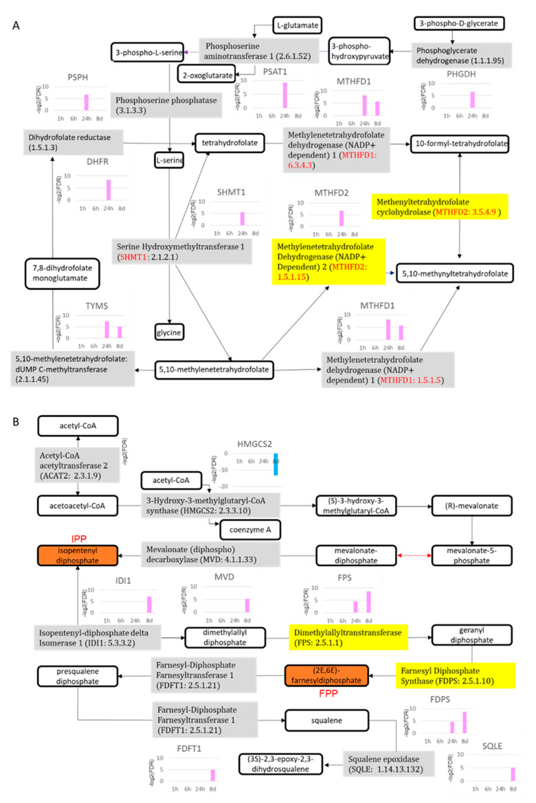
The Bhas 42 cell transformation assay (Bhas 42 CTA) is the first Organization for Economic Cooperation and Development (OECD)-certificated method used as a specific tool for the detection of the cell-transformation potential of tumor-promoting compounds, including non-genotoxic carcinogens (NGTxCs), as separate from genotoxic carcinogens. This assay offers the great advantage of enabling the phenotypic detection of oncotransformation. A key benefit of using the Bhas 42 CTA in the study of the cell-transformation mechanisms of tumor-promoting compounds, including non-genotoxic carcinogens, is that the cell-transformation potential of the chemical can be detected directly without treatment with a tumor-initiating compound since Bhas 42 cell line was established by transfecting the v-Ha-ras gene into a mouse fibroblast cloned cell line. Here, we analyzed the gene expression over time, using DNA microarrays, in Bhas 42 cells treated with the tumor-promoting compound 12-O-tetradecanoylphorbol-13-acetate (TPA), and NGTxC, with a total of three repeat experiments. This is the first paper to report on gene expression over time during the process of cell transformation with only a tumor-promoting compound. Pathways that were activated or inactivated during the process of cell transformation in the Bhas 42 cells treated with TPA were related not only directly to RAS but also to various pathways in the hallmarks of cancer.

Keywords: 12-O-tetradecanoylphorbol-13-acetate; Bhas 42 cells; cell-transformation assay; hallmarks of cancer; non-genotoxic carcinogen; over time; transcriptomics.


Conflict of interest statement

The authors declare no conflict of interest.

Figures

Figure 1
Figure 1
Morphological changes of Bhas 42 cells after treatment with TPA. (A) Solvent control, 1 h; (B) solvent control, 6 h; (C) solvent control, 24 h; (D) solvent control, 8 days; (E) TPA, 1 h; (F) TPA, 6 h; (G) TPA, 24 h; (H) TPA, 8 days.
Figure 2
Figure 2
Genes showing up (Up)-or down (Down)-regulation in response to TPA treatment. FDR, false-discovery rate.
Figure 3
Figure 3
Gene Ontology terms for selected genes. The GO terms up-regulated due to TPA treatment for (A) 1 h and 6 h. Gene Ontology terms for selected genes (continued). The GO terms up-regulated due to TPA treatment for (B) 24 h and 8 days.
Figure 3
Figure 3
Gene Ontology terms for selected genes. The GO terms up-regulated due to TPA treatment for (A) 1 h and 6 h. Gene Ontology terms for selected genes (continued). The GO terms up-regulated due to TPA treatment for (B) 24 h and 8 days.
Figure 4
Figure 4
Summary of Gene Ontology terms in chronological order.
Figure 5
Figure 5
The top-25 canonical pathways. TPA treatment for (A) 1 h and (B) 6 h. The line graph shows a list of genes whose expression increased or decreased at each treatment time and the −log value (p-value) obtained by testing a list of genes of the canonical pathway via Fisher’s exact test. The bar graph shows the rate (%) of the number of genes that were up-regulated (red) and the number of genes that were down-regulated (blue) when the number of genes in a known pathway was 100%. The top-25 canonical pathways (continued). TPA treatment for (C) 24 h and (D) 8 days. Other explanations are the same as for the graphs of the 1 h treatment and the 6 h treatment.
Figure 5
Figure 5
The top-25 canonical pathways. TPA treatment for (A) 1 h and (B) 6 h. The line graph shows a list of genes whose expression increased or decreased at each treatment time and the −log value (p-value) obtained by testing a list of genes of the canonical pathway via Fisher’s exact test. The bar graph shows the rate (%) of the number of genes that were up-regulated (red) and the number of genes that were down-regulated (blue) when the number of genes in a known pathway was 100%. The top-25 canonical pathways (continued). TPA treatment for (C) 24 h and (D) 8 days. Other explanations are the same as for the graphs of the 1 h treatment and the 6 h treatment.
Figure 6
Figure 6
Overlapping of the top-25 canonical pathways. TPA treatment for (A) 24 h and (B) 8 days. The color of the pathway is red when the z-score is larger than 0, and greenish when it is below 0. When the z-score is 0, the color is distinguished by the brightness of the monotone according to the value of the −log (p-value). Common genes among the respective pathways are connected by lines. The pathway connected by the blue line in (A) is the pathway of folate metabolism. The pathway connected by the blue line in (B) is the pathway for the biosynthesis of cholesterol.
Figure 7
Figure 7
Folate metabolic and cholesterol biosynthetic pathways. (A) Folate metabolic pathway; (B) cholesterol biosynthetic pathway. Gray and yellow are enzymes; others are biological products. 8d: 8 days.
Figure 8
Figure 8
Graphical summary of the molecule and its function following treatment with TPA for 24 h. Graphical summary was analyzed using IPA software. The activated molecule or function has a positive z-score and is therefore orange. The suppressed molecule or function has a negative z-score and is therefore blue. Estimated edges are shown as dotted lines (not solid lines).

Similar articles

Cited by

References

    1. Morita T., Hamada S., Masumura K., Wakata A., Maniwa J., Takasawa H., Yasunaga K., Hashizumef T., Honma M. Evaluation of the sensitivity and specificity of in vivo erythrocyte micronucleus and transgenic rodent gene mutation tests to detect rodent carcinogens. Mutat. Res. Genet. Toxicol. Environ. Mutagen. 2016;802:1–29. doi: 10.1016/j.mrgentox.2016.03.008. - DOI - PubMed
    1. Combes R.D. Detection of non-genotoxic carcinogens: Major barriers to replacement of the rodent assays. In: Van Zutphen L.F.M., Balls M., editors. Proceedings of the 2nd World Congress on Alternatives and Animal Use in the Life Sciences; Utrecht, The Netherlands. 20–24 October 1996; Amsterdam, The Netherlands: Elsevier; 1997. pp. 627–634.
    1. Ohmori K., Sasaki K., Asada S., Tanaka N., Umeda M. An assay method for the prediction of tumor promoting potential of chemicals by the use of Bhas 42 cells. Mutat. Res. 2004;557:191–202. doi: 10.1016/j.mrgentox.2003.10.014. - DOI - PubMed
    1. Sasaki K., Mizusawa H., Ishidate M. Isolation and characterization of ras-transfected BALB/3T3 clone showing morphological transformation by 12-O-tetradecanoyl-phorbol-13-acetate. Jpn. J. Cancer Res. 1988;79:921–930. doi: 10.1111/j.1349-7006.1988.tb00056.x. - DOI - PMC - PubMed
    1. Ohmori K., Umeda M., Tanaka N., Takagi T., Yoshimura I., Sasaki K., Asada S., Sakai A., Araki H., Asakura M., et al. An inter-laboratory collaborative study by the Non-Genotoxic Carcinogen Study Group in Japan, on a cell transformation assay for tumour promoters using Bhas 42 cells. Altern. Lab. Anim. 2005;33:619–639. doi: 10.1177/026119290503300616. - DOI - PubMed