. 2022 Mar 16;23(6):3221.
doi: 10.3390/ijms23063221.

Characterization of a Novel Aspect of Tissue Scarring Following Experimental Spinal Cord Injury and the Implantation of Bioengineered Type-I Collagen Scaffolds in the Adult Rat: Involvement of Perineurial-like Cells?

Affiliations

Characterization of a Novel Aspect of Tissue Scarring Following Experimental Spinal Cord Injury and the Implantation of Bioengineered Type-I Collagen Scaffolds in the Adult Rat: Involvement of Perineurial-like Cells?

Haktan Altinova et al. Int J Mol Sci. .

Abstract

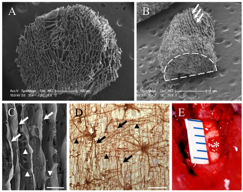
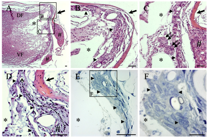
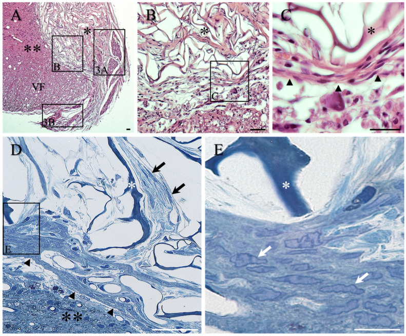
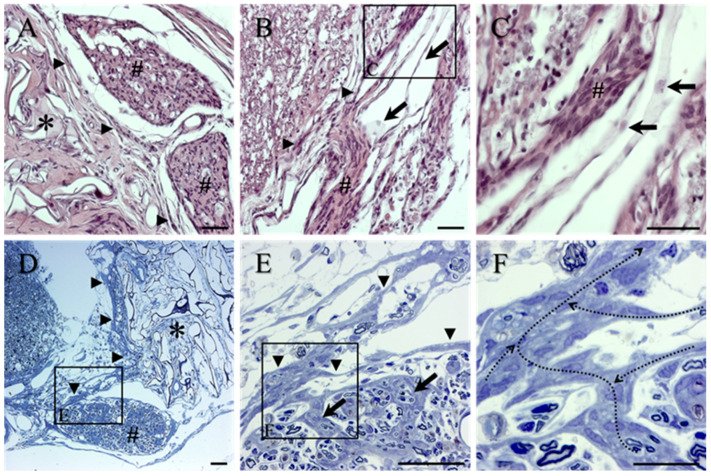
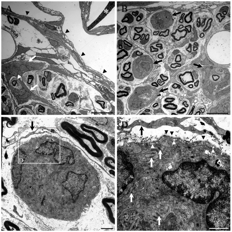
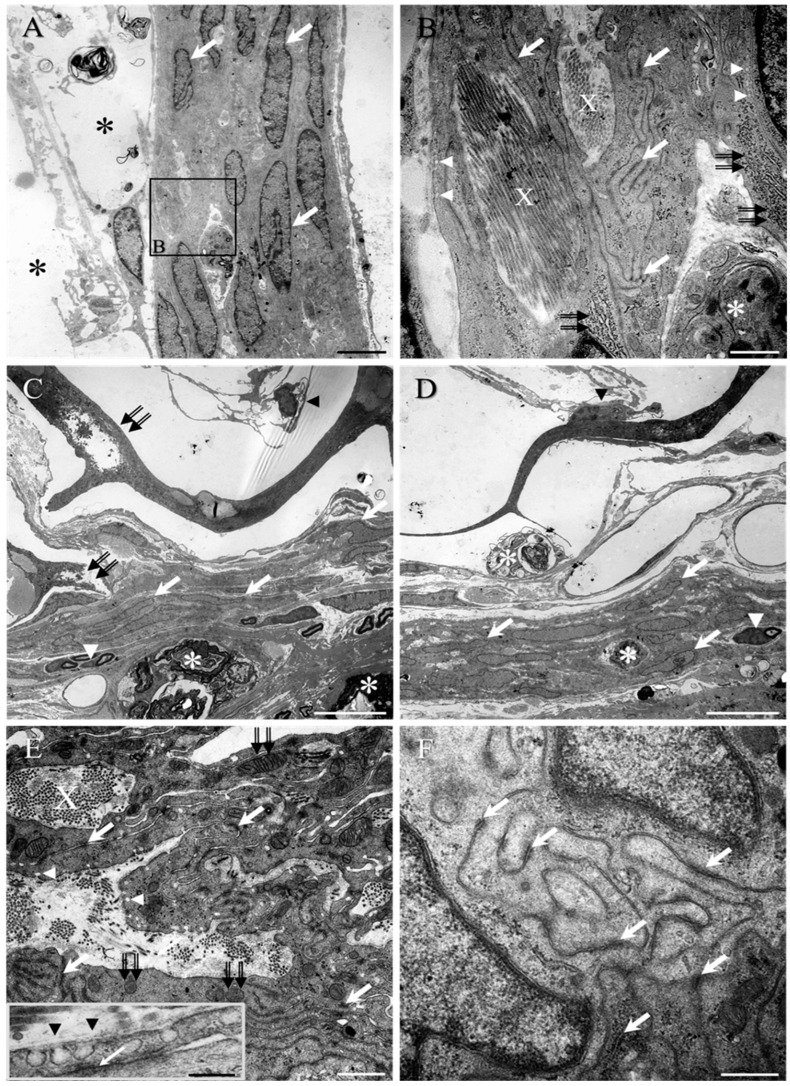
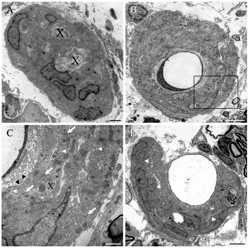
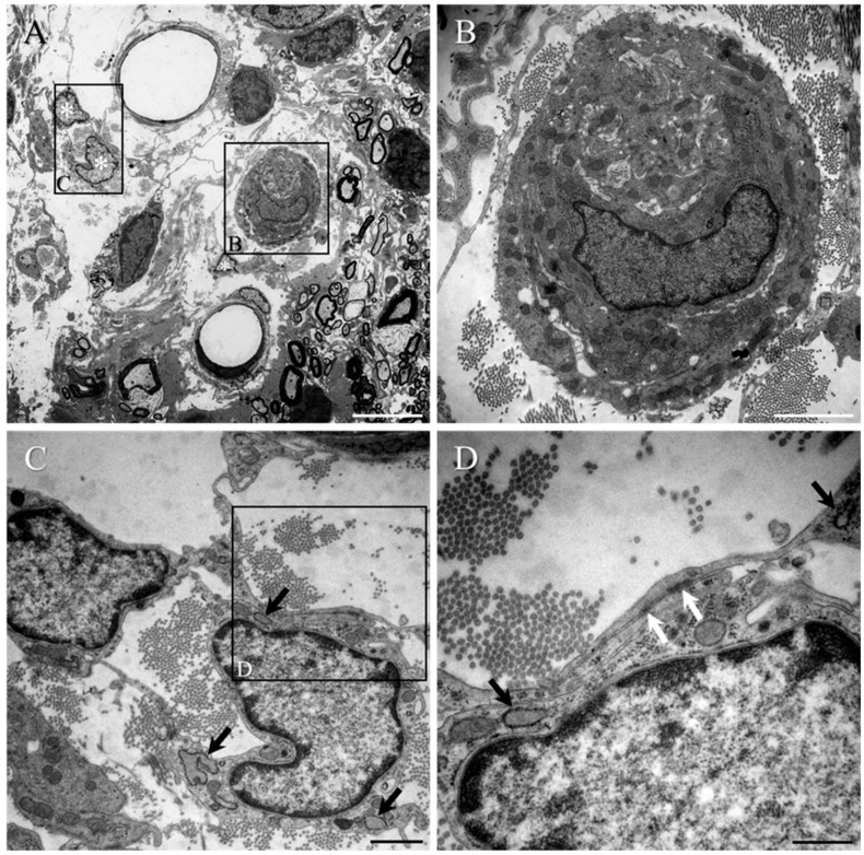
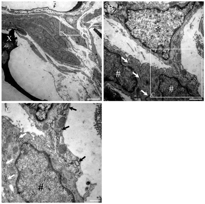
Numerous intervention strategies have been developed to promote functional tissue repair following experimental spinal cord injury (SCI), including the bridging of lesion-induced cystic cavities with bioengineered scaffolds. Integration between such implanted scaffolds and the lesioned host spinal cord is critical for supporting regenerative growth, but only moderate-to-low degrees of success have been reported. Light and electron microscopy were employed to better characterise the fibroadhesive scarring process taking place after implantation of a longitudinally microstructured type-I collagen scaffold into unilateral mid-cervical resection injuries of the adult rat spinal cord. At long survival times (10 weeks post-surgery), sheets of tightly packed cells (of uniform morphology) could be seen lining the inner surface of the repaired dura mater of lesion-only control animals, as well as forming a barrier along the implant-host interface of the scaffold-implanted animals. The highly uniform ultrastructural features of these scarring cells and their anatomical continuity with the local, reactive spinal nerve roots strongly suggest their identity to be perineurial-like cells. This novel aspect of the cellular composition of reactive spinal cord tissue highlights the increasingly complex nature of fibroadhesive scarring involved in traumatic injury, and particularly in response to the implantation of bioengineered collagen scaffolds.

Keywords: CNS-scarring; fibrotic encapsulation; implant interface; microstructured collagen scaffold; perineurial-like cells; spinal cord injury.


Conflict of interest statement

The authors declare no conflict of interest.

Figures

Figure 1
Figure 1
Biomimetic design of the microporous collagen scaffold. (A) SEM of the honeycomb-like appearance of the collagen scaffold cylinders after being removed from the 2 mm tissue punch (end-on view). (B) Tangential view of the scaffold hemi-cylinder generated by a mid-line incision along the axis of the cylinder. The profile of the end of the hemi-cylinder is highlighted by the dashed line. Longitudinal orientation of the porous framework can be identified on the scaffold surface (white arrows). (C) Higher-magnification SEM demonstrating the walls of longitudinally orientated scaffold framework (white arrows). The substantial fenestration between adjacent micropores is highlighted by the transversely orientated profiles (arrowheads). This bioengineered framework mimics, to some extent, the pattern and orientation of the astroglial framework of spinal cord white matter tracts as revealed by immunohistochemistry. (D) Glial fibrillary acidic protein (GFAP) immunohistochemistry of the interwoven pattern of longitudinal (black arrows) and transverse (black arrowheads) astrocytic processes in a longitudinal section of the lateral funiculus of the adult rat spinal cord. (E) A 2 mm long hemi-cylinder of the collagen scaffold (asterisk) fits neatly into the gap generated by the lateral funiculotomy of the adult rat cervical spinal cord, making excellent implant–host contact. The implant appears red after absorbing blood from the surrounding host tissue. The size of the lesion/implant is indicated by mm scale. Scale bars: A = 500 µm; B = 1 mm; C = 50 µm; D = 50 µm.
Figure 2
Figure 2
General morphology of lesion sites in lesion-only control animals, in transverse sections stained with H&E (AD) and toluidine blue semi-thin sections (E,F). (A) Low-magnification image of fluid-filled cystic cavities which were often divided by trabeculae (asterisks). Local spinal nerve roots are indicated by # and eosinophilic repaired dura mater is indicated by large arrow. For orientation, the dorsal funiculus is indicated by DF and the ventral funiculus by VF. (B) The lateral edge of cystic cavities (asterisks) was lined by layers of cells (arrowheads) directly at the medial surface of the repaired eosinophilic dura mater (arrow). (C) The described cell layers were observed to be in continuity with local, damaged spinal nerve roots (#; see also (A) for general overview). Reactive clusters of cells, possibly representing mini-fascicles of regenerating nervous tissue, can also be seen (double arrows). (D) The meandering black arrow indicates the arrangement of the reactive cell layer (arrowheads) medial to the eosinophilic dura mater (arrow). (E) The nuclei of the cells within these layers were better visualised in the toluidine blue-stained semi-thin sections, and displayed a fine rim of dense heterochromatin surrounding a medium-pale euchromatin (e.g., arrowheads; shown at higher magnification in (F)). Scale bars: AE = 50 µm; F = 20 µm.
Figure 3
Figure 3
Morphology of implanted collagen scaffold in transverse sections stained with H&E (AC) and for toluidine blue in semi-thin sections (D,E). (A) Low-magnification image of implanted scaffold (single asterisk) reveals the convoluted framework of the collagen scaffold and demonstrates the apparently good contact with the surrounding host spinal cord tissue (double asterisks). For orientation, the ventral funiculus is indicated by VF. Boxed area of the transition zone shown at higher magnification in Figure 2B. Other boxed areas shown at higher magnification in (A,B). (B) The presence of a conspicuous band of overlapping, elongated cells formed a transition zone at the interface between the collagen scaffold (asterisk) and the surrounding host tissue. Boxed area shown at higher magnification in (C). (C) Higher magnification of the band of the multiple overlapping cells (black arrowheads) coursing between the edge of the implant (asterisk) and the surrounding spinal cord tissue. (D) The morphology of the band of cells forming the transition zone around the implanted scaffold was more clearly seen in the toluidine blue semi-thin sections (black arrowheads). The darkly stained framework of the collagen scaffold (asterisk) highlights the open, porous nature of the scaffold with multiple, fine fibroblasts coursing amongst the palely stained collagen ECM deposits (black arrows). The overlapping cells and processes of the transition zone (arrowheads) separate the scaffold from the adjacent spinal cord parenchyma (double asterisk). (E) At high magnification, the uniform nuclear morphology of the tightly packed cells is apparent (white arrows). This observation is strikingly similar to the nuclear morphology of the cells in the lateral tissue bridge of the control group (compare with Figure 1F). Scale bars: AD = 50 µm; E = 20 µm.
Figure 4
Figure 4
Morphology of reactive spinal nerve roots in transverse spinal cord sections from the collagen scaffold-implanted group stained with H&E (AC) and for toluidine blue in semi-thin sections (DF). (A) Dorsal spinal nerve roots (#) located between thin sheets of overlapping, elongated reactive cells (arrowheads) covering the outer-most surface of the implanted collagen scaffold (asterisk) and the repaired dura mater. (B) Pale staining of the leptomeninges (arrows, also seen at higher magnification in (C)). (C) Higher magnification of the leptomeninges (arrows) is demonstrated close to a damaged spinal nerve rootlet (#). Note the morphological differences between leptomeningeal cells and the reactive cells of the rootlet (#). (D) Areas of thickened, reactive perineurium that circumscribed part of a damaged ventral nerve root could be seen extending towards and along the medial edge of the implanted scaffold (arrowheads). The porous framework of the scaffold (asterisk) contains abundant deposits of lightly stained ECM. (E) Higher magnification of the boxed area in (D). The damaged nerve root is surrounded by the overlapping cells and processes of reactive PNLC (arrowheads), which also formed the so-called septae within the damaged, reactive roots (arrows). (F) Higher magnification of the boxed area in (E). The continuity of the reactive cells that formed the intra-spinal nerve root septae with the cells of the surrounding perineurium is indicated by the meandering dotted arrows. Scale bars: AE = 50 µm; F = 20 µm.
Figure 5
Figure 5
Ultrastructure of the inter-fascicular septae and cell clusters in damaged spinal nerve roots of the collagen scaffold-implanted group. (A) Transmission electron microscopy demonstrated the reactive perineurial septae (e.g., white arrows) of a damaged/regenerated spinal nerve root that is close to the implanted collagen scaffold (asterisk). The perineurial surface of the nerve root is indicated by black arrowheads. This particular septum can be seen partially surrounding a loosely packed group of Schwann cell-myelinated axons as well as a single, isolated axon (white arrowhead). (B) The reactive PNLC also form rounded clusters or nests of cells (black arrows) that can even form part of the regenerated mini-fascicles and are loosely encircled by the fine processes of fibroblast-like cells. (C) Higher magnification of a cell cluster containing 3 PNLC nuclei. (D) High magnification of boxed area in (C). Many electron-dense, tight junctions can be seen between the intricately interwoven and overlapping processes of the reactive cells (white arrows), and numerous pinocytic caveolae (white arrowheads) are also evident. A discontinuous basal lamina (black arrowheads) is also present over the surface of the cluster. These ultrastructural features were all strikingly similar to those of perineurial cells. The fine overlapping processes of the fibroblast-like cells that surround the mini-fascicle appear to lack a basal lamina and tight junctions. Scale bars: A,B = 10 µm; C = 2 µm; D = 1 µm.
Figure 6
Figure 6
Ultrastructure of PNLC at the medial surface of the dura mater from the lesion-only control group (A,B) and at the transition zone from the collagen scaffold-implanted group (CF). (A) Multiple overlapping reactive PNLC forming the tissue bridge at the medial (or inner) edge of the repaired dura mater of lesion-only control animals. Cystic cavitation is indicated by asterisks. The similar directionality of the ovoid-shaped nuclei (white arrows) suggests that the cells had all adopted the same orientation. (B) Higher magnification of boxed area in (A). Bundles of collagen fibrils (X; see also within the transition zone of (E)) appear trapped within lacunae located between the cells, as do phagocytic macrophages (asterisk). Large numbers of electron-dense, tight junctions (white arrows; see also (E,F) for transition zone) are formed between the fine overlapping cell processes, and many pinocytotic vesicles can be seen close to the plasma membrane (white arrowheads; see also (E) for the transition zone). An abundance of rER (double black arrows) is also present within the cell body. (C,D) The same PNCL-like features, with predominantly ovoid-shaped nuclei, were observed in the transition zone around the implanted scaffolds (white arrows). The collagen framework of the scaffolds showed signs of degradation (e.g., double black arrows in (C)), with fibroblast-like cells and their long, fine processes being located within the lumen of the porous framework or adherent to the collagenous walls (black arrowheads in (C,D), respectively). Small groups of Schwann cell-myelinated axons (white arrowheads in (C,D), respectively) and phagocytic macrophages (asterisks in (C,D)) were also embedded amongst the overlapping cells and processes. (E,F) Pockets or lacunae of dense and loosely packed collagen fibrils (X) were trapped between the cell processes, which were connected by multiple electron-dense, tight junctions (white arrows in (E) and insert in (F)). Numerous pinocytotic vesicles (white arrowheads in (E); see also insert) and mitochondria (double arrows in (E)) suggested high levels of transport and metabolic activity. A discontinuous basal lamina was also regularly observed (e.g., black arrowheads, insert in (E)). Scale bars: A = 5 µm; B = 1 µm; C,D = 10 µm; E = 1 µm (insert, 200 nm); F = 500 nm.
Figure 7
Figure 7
Ultrastructure of PNLC clusters or nests close to the implant of the collagen scaffold-implanted group. (A) As typically seen with this population of reactive cells, isolated clusters or nests of reactive PNLC commonly included bundles of collagen fibrils within lacunae (X in (A,C)). (B) Some clusters or nests of PNLC were also observed to totally envelop capillaries within the damaged spinal cord parenchyma. (C) High magnification of boxed area in (B). Lacunae of trapped collagen (X) were scattered amongst the numerous overlapping processes with tight junctions (white arrows), pinocytic vesicles (white arrowheads), and discontinuous basal lamina (black arrowheads). (D) Some clusters only partially enveloped capillary walls. However, the nuclei of the PNLC (white arrowheads) appeared to be distinct when compared to that of a pericyte (asterisk) that had divested itself from the vessel wall. Scale bars: A = 2 µm; B,D = 5 µm; C = 1 µm.
Figure 8
Figure 8
(A) A small PNLC cluster and a pair of reactive pericytes can be seen within the heavily disorganised white matter that was located close to the transition zone of the collagen scaffold-implanted group. (B) High magnification of boxed area in (A). The cell cluster is surrounded by areas of densely packed collagen fibrils. The typical morphological appearance of the single PNLC nucleus (containing medium-dense euchromatin) is clearly different from that of the pericytes (containing much paler euchromatin) that have dissociated themselves from the local microcirculation (compare the nucleus in (B) with those in (C)). (C) High magnification of boxed area in (A). The cell body of the reactive pericyte contains relatively little cytoplasm but numerous free ribosomes and rER with conspicuously dilated cisternae (black arrows; see also (D)). (D) High magnification of boxed area in (C). The fine overlapping processes of the reactive pericytes displayed occasional electron-dense, tight junctions (white arrow). Scale bars: A = 5 µm; B = 2 µm; C = 1 µm; D = 500 nm.
Figure 9
Figure 9
Reactive pericytes at the transition zone of the collagen scaffold-implanted group. (A) A reactive pericyte (asterisk) can be seen in close contact to a capillary vessel wall that was located adjacent to the PNLC of the transition zone around the partially degraded collagen scaffold (X). (B) High magnification of boxed area in (A). The moderately electron-dense nuclei of the PNLC (indicated by #) were readily distinguishable from the paler, electron-lucent nucleus of the pericyte (asterisk), which could be seen extending towards and making contact with the PNLC. Electron-dense, tight junctions between the PNLC were detectable (white arrows), even at moderately low magnification. (C) High magnification of the boxed area in (B). The pericyte process, extending from the cell body, displayed the characteristically dilated rER (arrows) that was at least double the width of that of the PNLC. The process of the pericyte made close contact with the plasma membrane of PNLC, but no tight junctions could be observed. Scale bars: A = 5 µm; B = 1 µm; C = 500 nm.

Similar articles

Cited by

References

    1. Bunge R.P., Puckett W.R., Becerra J.L., Marcillo A., Quencer R.M. Observations on the pathology of human spinal cord injury. A review and classification of 22 new cases with details from a case of chronic cord compression with extensive focal demyelination. Adv. Neurol. 1993;59:75–89. - PubMed
    1. Kakulas B., Taylor J. Pathology of injuries of the vertebral column and spinal cord. Handb. Clin. Neurol. 1992;17:21–51.
    1. Silver J., Miller J.H. Regeneration beyond the glial scar. Nat. Rev. Neurosci. 2004;5:146–156. doi: 10.1038/nrn1326. - DOI - PubMed
    1. Fawcett J.W., Asher R.A. The glial scar and central nervous system repair. Brain Res. Bull. 1999;49:377–391. doi: 10.1016/S0361-9230(99)00072-6. - DOI - PubMed
    1. Stichel C.C., Muller H.W. The CNS lesion scar: New vistas on an old regeneration barrier. Cell Tissue Res. 1998;294:1–9. doi: 10.1007/s004410051151. - DOI - PubMed