. 2022 Mar 17;23(6):3243.
doi: 10.3390/ijms23063243.

Vector and Host C-Type Lectin Receptor (CLR)-Fc Fusion Proteins as a Cross-Species Comparative Approach to Screen for CLR-Rift Valley Fever Virus Interactions

Affiliations

Vector and Host C-Type Lectin Receptor (CLR)-Fc Fusion Proteins as a Cross-Species Comparative Approach to Screen for CLR-Rift Valley Fever Virus Interactions

Kathleen Schön et al. Int J Mol Sci. .

Abstract

Rift Valley fever virus (RVFV) is a mosquito-borne bunyavirus endemic to Africa and the Arabian Peninsula, which causes diseases in humans and livestock. C-type lectin receptors (CLRs) represent a superfamily of pattern recognition receptors that were reported to interact with diverse viruses and contribute to antiviral immune responses but may also act as attachment factors or entry receptors in diverse species. Human DC-SIGN and L-SIGN are known to interact with RVFV and to facilitate viral host cell entry, but the roles of further host and vector CLRs are still unknown. In this study, we present a CLR-Fc fusion protein library to screen RVFV-CLR interaction in a cross-species approach and identified novel murine, ovine, and Aedes aegypti RVFV candidate receptors. Furthermore, cross-species CLR binding studies enabled observations of the differences and similarities in binding preferences of RVFV between mammalian CLR homologues, as well as more distant vector/host CLRs.

Keywords: Aedes aegypti; C-type lectin domain-containing proteins; C-type lectin receptors; Rift Valley fever virus; fusion proteins.


Conflict of interest statement

The authors declare no conflict of interest.

Figures

Figure 1
Figure 1
Alignment of 28 C-type lectin receptor amino acid sequences (ae: Aedes aegypti, h: Homo sapiens sapiens, m: Mus musculus, o: Ovis aries). Amino acids similarity analysed with Score Matrix Blossum62 (Threshold 1) and symbolised in light grey (60–79% similarity), dark grey (80–99%), and black (100%). Hydrophobic part, indicating a transmembrane region, marked in dark red below the sequence. Above, consensus and mean pairwise identity over all amino acid sequences shown (green: 100% identity; yellow: 30–99%; red: <30%). Mosquito amino acid sequences were obtained from VectorBase and mammalian sequences from NCBI (Table 1 and Table 2). Sequence analysis was performed, and figures were prepared using the Geneious Prime software.
Figure 2
Figure 2
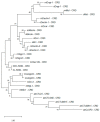
Maximum likelihood phylogenetic analysis of the carbohydrate recognition domain (CRD) of 28 C-type lectin receptors. Amino acid sequences of the respective CRDs were modelled by using bootstrap with 500 replications with the Jones–Taylor–Thornton plus frequency model and a discrete gamma distribution with five rate categories. Percentage frequencies from the bootstrap procedure are shown at each knot. Mosquito amino acid sequences were obtained from VectorBase, while mammalian sequences were from NCBI (Table 1 and Table 2). ae: Aedes aegypti, h: Homo sapiens sapiens, m: Mus musculus, o: Ovis aries. Phylogenetic analysis was performed with MEGA Software.
Figure 3
Figure 3
Mosquito CLR–hFc fusion proteins: (A) schematic overview of five Aedes aegypti transcripts (―) encoding for CTLDcps. Open reading frame is marked in grey, and nucleotides extracted for produced hFc–fusion proteins are highlighted in blue. Start codon and four conserved genetic motifs symbolised, as shown in the legend. AaedL5.1 CLR nucleotide sequences were obtained from VectorBase; (B) vector map of CLR domain ligated into pFUSE–hIgG1–Fc2 (Invitrogen); (C,D) Western blot image of anti-hFc stained mosquito (C) and ovine (D) CLR–hFc fusion proteins and hFc-empty control. Lane M: protein molecular standard, in kDa.
Figure 4
Figure 4
Mosquito CLR–hFc fusion protein interaction to mannan and zymosan: (A) ELISA-based screening of mannan with five mosquito CLR–hFc fusion proteins, in the presence and absence of EDTA. mDectin-2/oDectin-2 are known to recognise mannan (positive control) [35,41] and hFc-empty employed as negative control. One representative of n = 4 (EDTA n = 2) independent experiments shown; (B) ELISA-based screening of zymosan with five mosquito CLR–hFc fusion proteins, in the presence and absence of EDTA. mDectin-1/oDectin-1 are known to recognise zymosan (positive control) [35,43] and hFc-empty employed as negative control; (A,B) to discard possible false positives, the dotted line represents the cutoff defined as the threefold margin of the absorbance value relative to hFc, based on previous screenings with the CLR–hFc fusion protein library [34,46]. Data are depicted as mean + SEM of duplicates. One representative of n = 4 (EDTA n = 2) independent experiments is shown. One-way ANOVA, along with subsequent pairwise Tukey tests, was performed to compare the binding of the CLR–hFc fusion proteins above the threshold to PBS control and the EDTA supplementation each; **** p < 0.0001.
Figure 5
Figure 5
CLR–hFc fusion proteins across species bind to RVFV MP12: (A) ELISA-based binding study of purified RVFV MP12/Mock with murine CLR–hFc fusion proteins in the presence and absence of EDTA. hDC-SIGN served as positive control [24,45] and hFc-empty as negative control. One representative of n = 5 (EDTA: n = 2) independent experiments is shown; (B) ELISA-based screening of purified RVFV MP12/Mock with ovine CLR–hFc fusion proteins, in the presence and absence of EDTA. hDC-SIGN served as positive control and hFc-empty as negative control. One representative of n = 3 (EDTA: n = 2) independent experiments is shown; (C) ELISA-based screening of purified RVFV MP12/Mock with Aedes aegypti CLR–hFc fusion proteins, in the presence and absence of EDTA. hDC-SIGN served as positive control and hFc-empty as negative control. One representative of n = 3 (EDTA: n = 2) independent experiments is shown; (AC) to discard possible false positives, the dotted line represents the cutoff defined as the threefold margin of the absorbance value relative to hFc, based on previous screenings with the CLR–hFc fusion protein library [34,46]. Data are depicted as mean + SEM of duplicates. One-way ANOVA with subsequent pairwise Tukey tests were performed to compare the binding of the CLR–hFc fusion proteins above the threshold to mock and the EDTA supplementation; ** p < 0.01, *** p < 0.0005, **** p < 0.0001.

References

    1. Sukhralia S., Verma M., Gopirajan S., Dhanaraj P.S., Lal R., Mehla N., Kant C.R. From dengue to Zika: The wide spread of mosquito-borne arboviruses. Eur. J. Clin. Microbiol. Infect. Dis. 2019;38:3–14. doi: 10.1007/s10096-018-3375-7. - DOI - PubMed
    1. Girard M., Nelson C.B., Picot V., Gubler D.J. Arboviruses: A global public health threat. Vaccine. 2020;38:3989–3994. doi: 10.1016/j.vaccine.2020.04.011. - DOI - PMC - PubMed
    1. Kuhn J.H., Adkins S., Alioto D., Alkhovsky S.V., Amarasinghe G.K., Anthony S.J., Avšič-Županc T., Ayllón M.A., Bahl J., Balkema-Buschmann A., et al. 2020 taxonomic update for phylum Negarnaviricota (Riboviria: Orthornavirae), including the large orders Bunyavirales and Mononegavirales. Arch. Virol. 2020;165:3023–3072. doi: 10.1007/s00705-020-04731-2. - DOI - PMC - PubMed
    1. Walter C.T., Barr J.N. Recent advances in the molecular and cellular biology of bunyaviruses. J. Gen. Virol. 2011;92:2467–2484. doi: 10.1099/vir.0.035105-0. - DOI - PubMed
    1. Wright D., Kortekaas J., Bowden T.A., Warimwe G.M. Rift Valley fever: Biology and epidemiology. J. Gen. Virol. 2019;100:1187–1199. doi: 10.1099/jgv.0.001296. - DOI - PMC - PubMed

Substances

LinkOut - more resources