. 2022 Mar 17;23(6):3258.
doi: 10.3390/ijms23063258.

In Vitro Studies Regarding the Safety of Chitosan and Hyaluronic Acid-Based Nanohydrogels Containing Contrast Agents for Magnetic Resonance Imaging

Affiliations

In Vitro Studies Regarding the Safety of Chitosan and Hyaluronic Acid-Based Nanohydrogels Containing Contrast Agents for Magnetic Resonance Imaging

Cecilia Virginia Gheran et al. Int J Mol Sci. .

Abstract

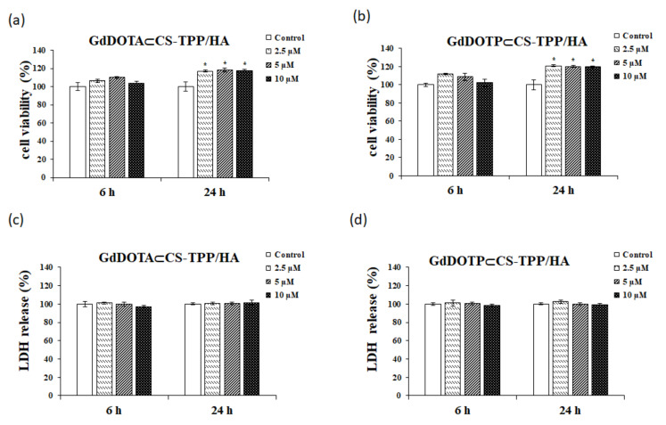
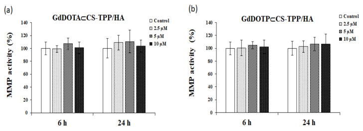
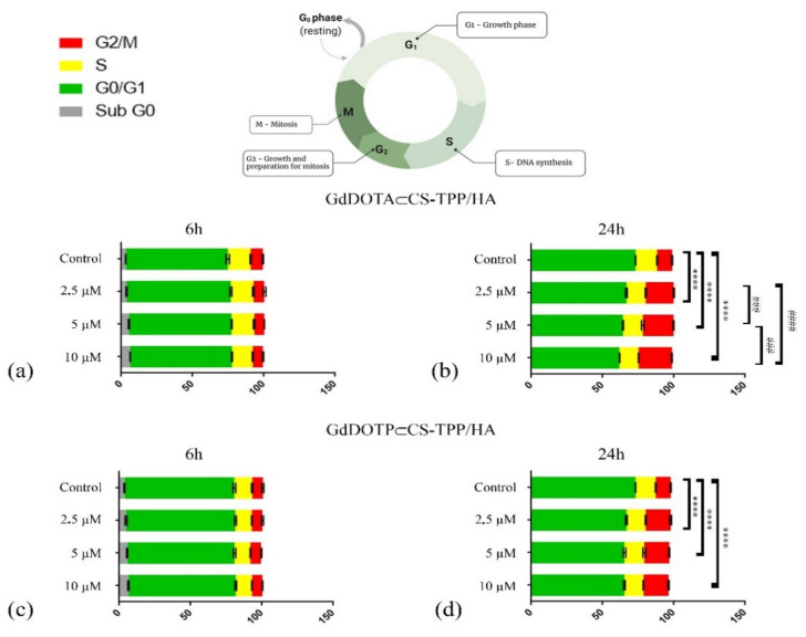
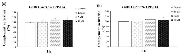
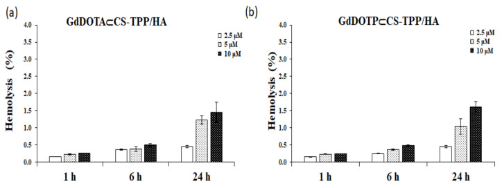
The aim of this study was to investigate the biocompatibility of contrast agents, such as gadolinium 1, 4, 7, 10 tetraazacyclo-dodecane tetraacetic acid (GdDOTA) and gadolinium dioctyl terephthalate (GdDOTP), encapsulated in a polymeric matrix containing chitosan and hyaluronic acid using RAW264.7 murine macrophages and human blood samples. The cell viability and cytotoxicity were evaluated by 3-(4,5-dimethylthiazol-2-yl)-2,5-diphenyltetrazolium bromide (MTT) and lactate dehydrogenase (LDH) assays, while cell cycle analysis was determined in RAW264.7 cells using flow cytometry. The mitochondrial membrane potential (MMP), hemolytic index, complement activation, and thrombogenic potential of gadolinium (Gd) containing nanohydrogels were measured by fluorometric and spectrophotometric methods. Taken together, our results demonstrate the good bio- and hemocompatibility of chitosan-based nanohydrogels with the RAW264.7 cell line and human blood cells, suggesting that these could be used as injectable formulations for the magnetic resonance imaging diagnostic of lymph nodes.

Keywords: RAW 264.7 cell line; biocompatibility; chitosan; contrast agents; hemocompatibility; nanohydrogel.


Conflict of interest statement

The authors declare no conflict of interest. The funders had no role in the design of the study; in the collection, analyses, or interpretation of data; in the writing of the manuscript, or in the decision to publish the results.

Figures

Figure 1
Figure 1
The MTT (a,b) and LDH (c,d) assays in presence of RAW 264.7 cells after 6- and 24 h exposed to doses of 2.5, 5 and 10 μM of (a,c) GdDOTA⊂CS-TPP/HA and (b,d) GdDOTP⊂CS-TPP/HA NGs. Untreated cells were used as a control. Data are expressed as mean ± SD (n = 3). The asterisks represent the statistical significance obtained by the Student’s test, as follows: * p < 0.05 (significant).
Figure 2
Figure 2
The level of mitochondrial membrane potential in RAW 264.7 murine macrophages cells after 6 -and 24 h of exposure to doses of 2.5, 5 and 10 μM of (a) GdDOTA⊂CS-TPP/HA and (b) GdDOTP⊂CS-TPP/HA NGs. Untreated cells were used as a control. Data are expressed as mean ± SD (n = 3).
Figure 3
Figure 3
Cell cycle distribution in RAW 267.4 murine macrophage cells treated with (a) GdDOTA⊂CS-TPP/HA NGs for 6 h; (b) GdDOTA⊂CS-TPP/HA NGs for 24 h: **** p < 0.0001 control versus 2.5 µM_G0/G1 and G2/M; control versus 5 µM_G0/G1 and G2/M and control versus 10 µM_G0/G1 and G2/M); ### p < 0.001 2.5 µM versus 5 µM _G0/G1 and G2/M and 5 µM versus 10 µM _G0/G1 and G2/M; #### p < 0.0001 2.5 µM versus 10 µM _G0/G1 and G2/M; (c) GdDOTP⊂CS-TPP/HA NGs for 6 h and (d) GdDOTP⊂CS-TPP/HA NGs for 24 h: **** p < 0.0001 control versus 2.5 µM_G0/G1 and G2/M; control versus 5 µM_G0/G1 and G2/M and control versus 10 µM_G0/G1 and G2/M.
Figure 4
Figure 4
Classical activation of the complement system after 1 h of exposure to doses of 2.5, 5 and 10 μM of (a) GdDOTA⊂CS-TPP/HA and (b) GdDOTP⊂CS-TPP/HA NGs. A human serum sample incubated at 37 °C served as a negative (normal) control. Data are expressed as mean ± SD (n = 3).
Figure 5
Figure 5
The percentage of hemolysis after 1, 6 and 24 h of incubation with (a) GdDOTA⊂CS-TPP/HA and (b) GdDOTP⊂CS-TPP/HA NGs, at doses of 2.5, 5 and 10 μM. Data are expressed as mean ± SD (n = 3). As a positive (100% hemolysis) and negative (0% hemolysis) controls, supernatants resulting from red blood cells treated with Triton X-100 1% and physiological serum (0.9% NaCl) respectively have been used.

Similar articles

Cited by

References

    1. Wunderbaldinger P. Problems and prospects of modern lymph node imaging. Eur. J. Radiol. 2006;58:325–337. doi: 10.1016/j.ejrad.2005.12.037. - DOI - PubMed
    1. Luo D., Shan Z., Liu Q., Cai S., Ma Y., Li Q., Li X. The correlation between tumor size, lymph node status, distant metastases and mortality in rectal cancer patients without neoadjuvant therapy. J. Cancer. 2021;12:1616–1622. doi: 10.7150/jca.52165. - DOI - PMC - PubMed
    1. Van den Brekel M.W. Lymph node metastases: CT and MRI. Eur. J. Radiol. 2000;33:230–238. doi: 10.1016/S0720-048X(99)00145-X. - DOI - PubMed
    1. Yanzhang W., Guanghua L., Zhihao Z., Zhixiong W., Zhao W. The risk of lymph node metastasis in gastric cancer conforming to indications of endoscopic resection and pylorus-preserving gastrectomy: A single-center retrospective study. BMC Cancer. 2021;21:1280. doi: 10.1186/s12885-021-09008-8. - DOI - PMC - PubMed
    1. Misselwitz B. MR contrast agents in lymph node imaging. Eur. J. Radiol. 2006;58:375–382. doi: 10.1016/j.ejrad.2005.12.044. - DOI - PubMed

LinkOut - more resources