Review
. 2022 Mar 17;23(6):3265.
doi: 10.3390/ijms23063265.

Polymeric Hydrogels for In Vitro 3D Ovarian Cancer Modeling

Affiliations
Review

Polymeric Hydrogels for In Vitro 3D Ovarian Cancer Modeling

Simona Braccini et al. Int J Mol Sci. .

Abstract

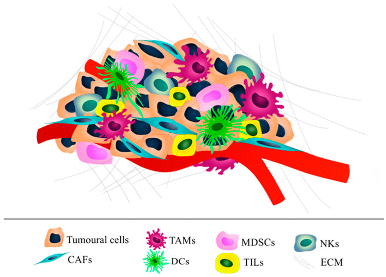
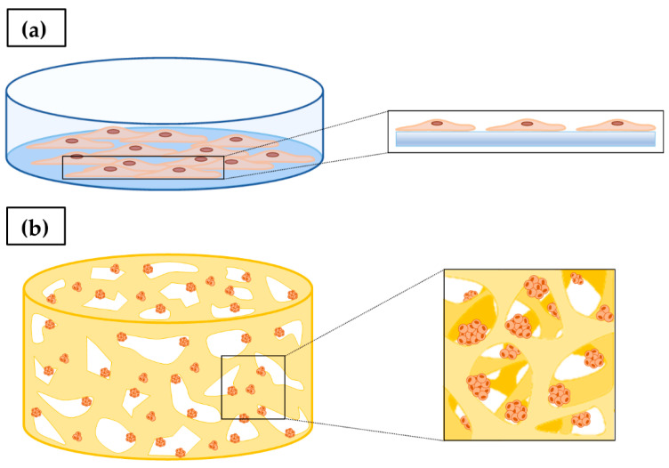
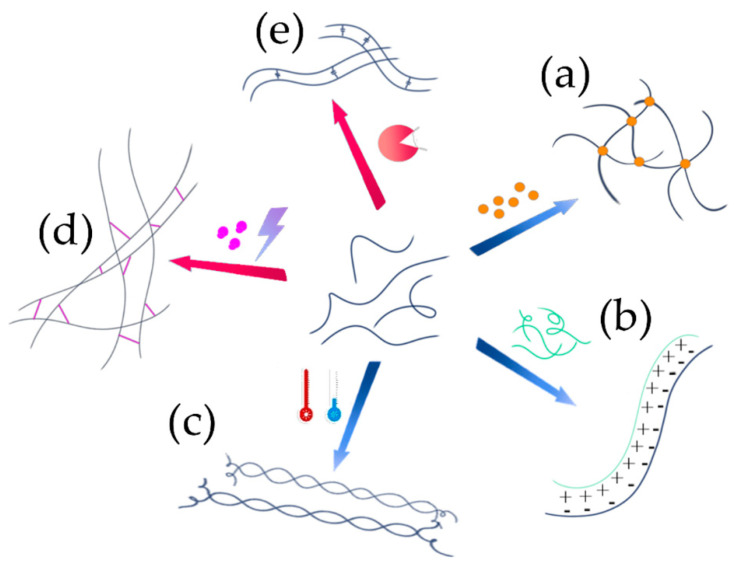
Ovarian cancer (OC) grows and interacts constantly with a complex microenvironment, in which immune cells, fibroblasts, blood vessels, signal molecules and the extracellular matrix (ECM) coexist. This heterogeneous environment provides structural and biochemical support to the surrounding cells and undergoes constant and dynamic remodeling that actively promotes tumor initiation, progression, and metastasis. Despite the fact that traditional 2D cell culture systems have led to relevant medical advances in cancer research, 3D cell culture models could open new possibilities for the development of an in vitro tumor microenvironment more closely reproducing that observed in vivo. The implementation of materials science and technology into cancer research has enabled significant progress in the study of cancer progression and drug screening, through the development of polymeric scaffold-based 3D models closely recapitulating the physiopathological features of native tumor tissue. This article provides an overview of state-of-the-art in vitro tumor models with a particular focus on 3D OC cell culture in pre-clinical studies. The most representative OC models described in the literature are presented with a focus on hydrogel-based scaffolds, which guarantee soft tissue-like physical properties as well as a suitable 3D microenvironment for cell growth. Hydrogel-forming polymers of either natural or synthetic origin investigated in this context are described by highlighting their source of extraction, physical-chemical properties, and application for 3D ovarian cancer cell culture.

Keywords: 3D cell culture; hydrogel; ovarian cancer; polymer; scaffold.


Conflict of interest statement

The authors declare no conflict of interest.

Figures

Figure 1
Figure 1
Total number of publications per year with “3D culture” topic. Source: Web of Science: Science Citation Index Expanded [13].
Figure 2
Figure 2
Graphical representation of ovarian tumor microenvironment (TME). In particular, the following components are represented: tumoral cells, cancer-associated fibroblasts (CAFs), tumor-associated macrophages (TAMs), dendritic cells (DCs), myeloid-derived suppressor cells (MDSCs), tumor-infiltrating lymphocytes (TILs), natural killers (NKs) and ECM structural elements.
Figure 3
Figure 3
(a) Two-dimensional cell culture: cells cultured in a petri dish arranged to form a cellular monolayer with a flat cell shape that does not closely recapitulate the real physiological cell morphology. (b) 3D cell culture: in the 3D microenvironment, cells form multicellular aggregates, presenting a morphology and behavior more representative of in vivo systems.
Figure 4
Figure 4
Graphical representation of hydrogel formation process through physical (blue arrows) or chemical (red arrows) interactions among macromolecular chains. (a) Ionic crosslinking, (b) polyelectrolyte complex (PEC) formation, (c) thermoresponsive gelling process, (d) photoactivated crosslinking, and (e) enzymatic crosslinking.
Figure 5
Figure 5
The cell adhesion process on scaffold surface. (a) The cell comes in contact with scaffold surface and loosely attaches onto the substrate, (b) the cell starts to flatten, (c) the cell spread its membrane and form focal adhesions that connect the cell securely on the scaffold surface with intracellular actin filaments (stress fibers) through integrin, and (d) the cell begins to migrate to the scaffold surface generating new EMC (the green arrow indicates migration direction).
Figure 6
Figure 6
Schematic representation of scaffold fabrication techniques. (a) Freeze-drying, (b) supercritical drying, (c) solvent casting, (d) phase separation, (e) gas foaming, (f) electrospinning, and (g) additive manufacturing.
Figure 7
Figure 7
Hydrogel-based scaffolds by additive manufacturing (AM). (a) Top view of 3D bio-printed construct based on GelMA [192], (b) photographic image of ten-layer printed scaffold with Ink H4-RGD [193] and, (c) representative photograph of a chitosan/γ-PGA PEC hydrogel and representative confocal laser scanning microscopy micrograph of BxPC-3 cells grown on the PEC hydrogel [97].
Figure 8
Figure 8
Chemical structure of polysaccharides investigated for hydrogel-based scaffolds in OC modeling. (a) Chitosan obtained by chitin denaturation, (b) cellulose, (c) alginate, and (d) agarose.
Figure 9
Figure 9
Chitosan and cellulose hydrogels. (a) Chemical structure of sChi (green)-oxAlg (blue) unit, and schematic representation of cell seeding process in U-bottom 96-well plates; (b) setup for automated seeding of cells via Tecan Freedom EVO liquid handling station; (c) cell-seeded scaffolds at day 1 (scale bar = 10 mm) [197]. (d) SEM morphology of BC and BC-Chi scaffolds; (e) A2780 cell proliferation on BC and BC-Chi scaffolds for 1, 3, 5, and 7 days [198].
Figure 10
Figure 10
Agarose hydrogel. (a) SKOV3 cell proliferation in 3D and 2D cultures. */#, **/##, and ***/### denote p < 0.05, p < 0.01, and p < 0.001, respectively. (b) Live/dead cell assay: (i) 2D control, (ii) 3D control, and (iii) 3D agarose. Scale bar is 100 µm [199].
Figure 11
Figure 11
Chemical structure of proteins investigated for hydrogel-based scaffolds in OC modeling. (a) Triple chain structure of collagen fibrils and chemical structure of the most common tripeptide sequence found in collagen, composed of Gly, Pro and Hyp sequences, (b) gelatin and its reaction with methacrylic anhydride to form photocrosslinkable gelatin–methacryloyl (GelMA).
Figure 12
Figure 12
Collagen hydrogels. (a) SEM micrographs of collagen mesh (right) and OV-NC cells in collagen scaffolds (left) (yellow arrows indicate OV-NC cell clusters) [202]. (b) Jellyfish collagen-based sponges molded on 96-well plate (diameter of 5 mm), (c) SEM micrograph of scaffold porous structure, and (d) cell proliferation and migration throughout 3D collagen scaffold cross-section [203].
Figure 13
Figure 13
Gelatin–methacryloyl (GelMA) hydrogels. (a) GelMA-based hydrogel preparation: gelatin (1) and methacrylic anhydride reacted to form GelMA (2); GelMA is dissolved in PBS at 37 °C and mixed with the photoinitiator and cells (3); the cross-linking reaction is induced by UV light (4); the hydrogel is cut into smaller units (5). (b) Representative confocal laser scanning microscopy micrographs of cells embedded within GelMA hydrogels obtained from various polymer concentrations (w/v) and relevant stiffness (2.5%, 0.7 kPa; 5%, 3.4 kPa; 7%, 7.3 kPa and 10%, 16.5 kPa); nuclei are stained in blue and actin cytoskeleton in red (scale bars, 100 μm) [204].
Figure 14
Figure 14
Chemical structure of synthetic polymers employed in hydrogel-based scaffolds for in vitro OC modeling. (a) Tetrapeptide sequence found in RIPA16-I peptide, and (b) poly(ethylene glycol) (PEG).
Figure 15
Figure 15
RADA16-I hydrogel. (a) Negatively stained TEM images showing the nanofiber morphology of RADA16-I (i,iv), Matrigel (ii,v), and collagen I (iii,vi) (scale bar, top panels (i–iii): ×8000; bottom panels (iv–vi): ×15,000). (b) Cisplatin and paclitaxel responses of chemosensitivity assay in HO-8910PM cells cultured in gel-cell clumps and common 2D flat cell plates: a (p < 0.01) compared with that of 2D cell culture model; b (p < 0.01) compared with that of Matrigel; c (p < 0.01) compared with that of RADA16-I hydrogel [206].
Figure 16
Figure 16
PEG-based hydrogel. (a) Schematic illustration of OC cells grown in a monolayer on traditional plastic surfaces (left) and in 3D as spheroids embedded within hydrogels (right). (b) OV-MZ-6 and SKOV-3 cells formed in 2D typical monolayers (left) and 3D systems (right), shown by phase contrast (top panel) and confocal (bottom panel; cell actin filaments stained with rhodamine phalloidin, nuclei with DAPI) microscopy images (scale bars, 75 μm) [208].

Similar articles

Cited by

References

    1. Stewart C., Ralyea C., Lockwood S. Ovarian Cancer: An Integrated Review. Semin. Oncol. Nurs. 2019;35:151–156. doi: 10.1016/j.soncn.2019.02.001. - DOI - PubMed
    1. National Cancer Institute Surveillance, Epidemiology, and End Results Program. Cancer Stat Facts: Ovarian Cancer. [(accessed on 15 February 2022)]; Available online: https://seer.cancer.gov.
    1. Lheureux S., Gourley C., Vergote I., Oza A.M. Epithelial ovarian cancer. Lancet. 2019;393:1240–1253. doi: 10.1016/S0140-6736(18)32552-2. - DOI - PubMed
    1. Cortez A.J., Tudrej P., Kujawa K.A., Lisowska K.M. Advances in ovarian cancer therapy. Cancer Chemother. Pharmacol. 2018;81:17–38. doi: 10.1007/s00280-017-3501-8. - DOI - PMC - PubMed
    1. Langhans S.A. Three-Dimensional in Vitro Cell Culture Models in Drug Discovery and Drug Repositioning. Front. Pharmacol. 2018;9:6. doi: 10.3389/fphar.2018.00006. - DOI - PMC - PubMed