. 2022 Mar 19;23(6):3324.
doi: 10.3390/ijms23063324.

The Prognostic Relevance of PMCA4 Expression in Melanoma: Gender Specificity and Implications for Immune Checkpoint Inhibition

Affiliations

The Prognostic Relevance of PMCA4 Expression in Melanoma: Gender Specificity and Implications for Immune Checkpoint Inhibition

Luca Hegedüs et al. Int J Mol Sci. .

Abstract

PMCA4 is a critical regulator of Ca2+ homeostasis in mammalian cells. While its biological and prognostic relevance in several cancer types has already been demonstrated, only preclinical investigations suggested a metastasis suppressor function in melanoma. Therefore, we studied the expression pattern of PMCA4 in human skin, nevus, as well as in primary and metastatic melanoma using immunohistochemistry. Furthermore, we analyzed the prognostic power of PMCA4 mRNA levels in cutaneous melanoma both at the non-metastatic stage as well as after PD-1 blockade in advanced disease. PMCA4 localizes to the plasma membrane in a differentiation dependent manner in human skin and mucosa, while nevus cells showed no plasma membrane staining. In contrast, primary cutaneous, choroidal and conjunctival melanoma cells showed specific plasma membrane localization of PMCA4 with a wide range of intensities. Analyzing the TCGA cohort, PMCA4 mRNA levels showed a gender specific prognostic impact in stage I-III melanoma. Female patients with high transcript levels had a significantly longer progression-free survival. Melanoma cell specific PMCA4 protein expression is associated with anaplasticity in melanoma lung metastasis but had no impact on survival after lung metastasectomy. Importantly, high PMCA4 transcript levels derived from RNA-seq of cutaneous melanoma are associated with significantly longer overall survival after PD-1 blockade. In summary, we demonstrated that human melanoma cells express PMCA4 and PMCA4 transcript levels carry prognostic information in a gender specific manner.

Keywords: immune checkpoint inhibition; lung metastasis; melanoma; plasma membrane calcium ATPase 4.


Conflict of interest statement

E. Livingstone has had intermittent advisory board relationships with Roche, BMS, Novartis, Sanofi and Actelion and has received travel grants and honoraria from Roche, BMS, MSD, Amgen, Pierre Fabre, SunPharma, Novartis, Boehringer-Ingelheim, medac. Dirk Schadendorf received research funding from Novartis and Bristol-Myers Squibb, served as consultant or/and has received honoraria from Amgen, Leo Pharma, Roche, Bristol-Myers Squibb, Merck Sharp & Dohme, Novartis, Incyte, Regeneron, 4SC, AstraZeneca, Immunocore, Pierre Fabre, Merck EMD, Pfizer, Philogen, Sanofi, Inflarx, Neracare and Array and travel support from Novartis, Bristol-Myers Squibb, Merck Sharp & Dohme, Roche and Pierre Fabre. All other authors declare no conflict of interest. AZ: received travel support from Novartis, Sanofi Grenzyme, Sun Pharma, and Bristol-Myers Squibb, outside the submitted work.

Figures

Figure 1
Figure 1
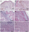
PMCA4b expression in human skin and mucosa. (A,B) In the skin, PMCA4b expression is increasing from the basal cells to the stratum spinosum and granulosum. The PMCA4b staining pattern resembles desmosomal localization. (C) In the vaginal mucosa, basal cells have the highest PMCA4b expression. (DF) The hematoxylin-eosin staining of the vulval epidermis shows a high number of pigment containing cells. (E) Melan-A staining (red) identifies the melanocytes in the basal cell layer that are characterized by a distinct nuclear morphology. (F) The plasma membrane between melanocytes and basal cells stained for PMCA4, whether the proteins are localized on the melanocytic plasma membrane as well cannot be unequivocally determined. Scale bars: (A,C): 100 µm; (B,DF): 10 µm.
Figure 2
Figure 2
PMCA4b expression in human nevus. No plasma membrane specific staining was observed in any of the nevi (A—junctional, B—compound, C—intradermal and D—dysplastic). Scale bars: (A) 100; µm; (BD): 50 µm.
Figure 3
Figure 3
Representative images of PMCA4b expression in human primary melanoma. (A) Weak but clear plasma membrane-specific PMCA4b expression in cutaneous melanoma. (B,C) Intermediate staining intensity in epitheloid (B) and mixed morphology (C). (D) High PMCA4b expression in a cutaneous melanoma with spindle cell morphology. (E,F) Plasma membrane specific PMCA4b expression in conjunctival (E) and choroidal (F) melanoma. Scale bars: (AD): 100 µm; (E,F): 50 µm.
Figure 4
Figure 4
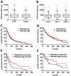
PMCA4 is a gender specific prognostic factor in the TCGA cohort of non-metastatic cutaneous melanoma patients. (A) There is no difference in the distribution of PMCA4 mRNA levels between male and female patients. (B) PMCA4 mRNA levels are independent of disease stage. (C,D) PMCA4 levels show no association with progression-free (C) or overall (D) survival in the overall cohort. (E) In female patients, high PMCA4 levels confer a significantly longer progression-free survival (p = 0.0037). (F) The difference in overall survival in female patients between high and low PMCA4 mRNA levels did not reach statistical significance (p = 0.0717).
Figure 5
Figure 5
PMCA4b expression in melanoma lung metastasis. (A) Weak but clear plasma membrane-specific PMCA4b staining in a lung metastasis. (B) Intermediate intensity staining. (C,D) High PMCA4b expression in epitheloid (C) and spindle cell (D) melanoma. Scale bar: (AC): 100 µm; (D): 200 µm.
Figure 6
Figure 6
Clinicopathological analysis of PMCA4b protein levels in melanoma lung metastasis. (A) There is no difference in the distribution of PMCA4b expression between male and female patients. (B) BRAF and NRAS mutation has no impact on PMCA4b staining intensity. (C,D) Melanoma specimens with anaplastic components showed decreased PMCA4b expression. (D) There is no prognostic impact of melanoma cell specific PMCA4b expression for overall survival after pulmonary metastasectomy (p = 0.437).
Figure 7
Figure 7
PMCA4 is a prognostic factor in PD-1 inhibitor treated melanoma patients. (A) There is no difference in the distribution of PMCA4 transcript levels between male (n = 71) and female (n = 50) patients (TPM—transcript per million). (B) There is no significant difference in PMCA4 mRNA levels between BRAF mutant (n = 51), NRAS mutant (n = 34) and double wild type (n = 36) tumors (C) PMCA4 levels in responders (n = 65) and in patients with progressive disease (n = 56; p = 0.0677). (D) PMCA4 levels showed no significant impact on progression-free survival after PD-1 blockade. (E) High PMCA4 transcript levels were associated with longer overall survival (p = 0.0182).

Similar articles

Cited by

References

    1. Siegel R.L., Miller K.D., Fuchs H.E., Jemal A. Cancer Statistics, 2021. CA Cancer J. Clin. 2021;71:7–33. doi: 10.3322/caac.21654. - DOI - PubMed
    1. Eddy K., Shah R., Chen S. Decoding Melanoma Development and Progression: Identification of Therapeutic Vulnerabilities. Front. Oncol. 2020;10:626129. doi: 10.3389/fonc.2020.626129. - DOI - PMC - PubMed
    1. Yeh I., von Deimling A., Bastian B.C. Clonal BRAF mutations in melanocytic nevi and initiating role of BRAF in melanocytic neoplasia. J. Natl. Cancer Inst. 2013;105:917–919. doi: 10.1093/jnci/djt119. - DOI - PMC - PubMed
    1. Shain A.H., Yeh I., Kovalyshyn I., Sriharan A., Talevich E., Gagnon A., Dummer R., North J., Pincus L., Ruben B., et al. The Genetic Evolution of Melanoma from Precursor Lesions. N. Engl. J. Med. 2015;373:1926–1936. doi: 10.1056/NEJMoa1502583. - DOI - PubMed
    1. Curtin J.A., Fridlyand J., Kageshita T., Patel H.N., Busam K.J., Kutzner H., Cho K.H., Aiba S., Brocker E.B., LeBoit P.E., et al. Distinct sets of genetic alterations in melanoma. N. Engl. J. Med. 2005;353:2135–2147. doi: 10.1056/NEJMoa050092. - DOI - PubMed

Substances