

Theoretical Studies of Cyanophycin Dipeptides as Inhibitors of Tyrosinases
- PMID: 35328756
- PMCID: PMC8950311
- DOI: 10.3390/ijms23063335
Theoretical Studies of Cyanophycin Dipeptides as Inhibitors of Tyrosinases
Abstract
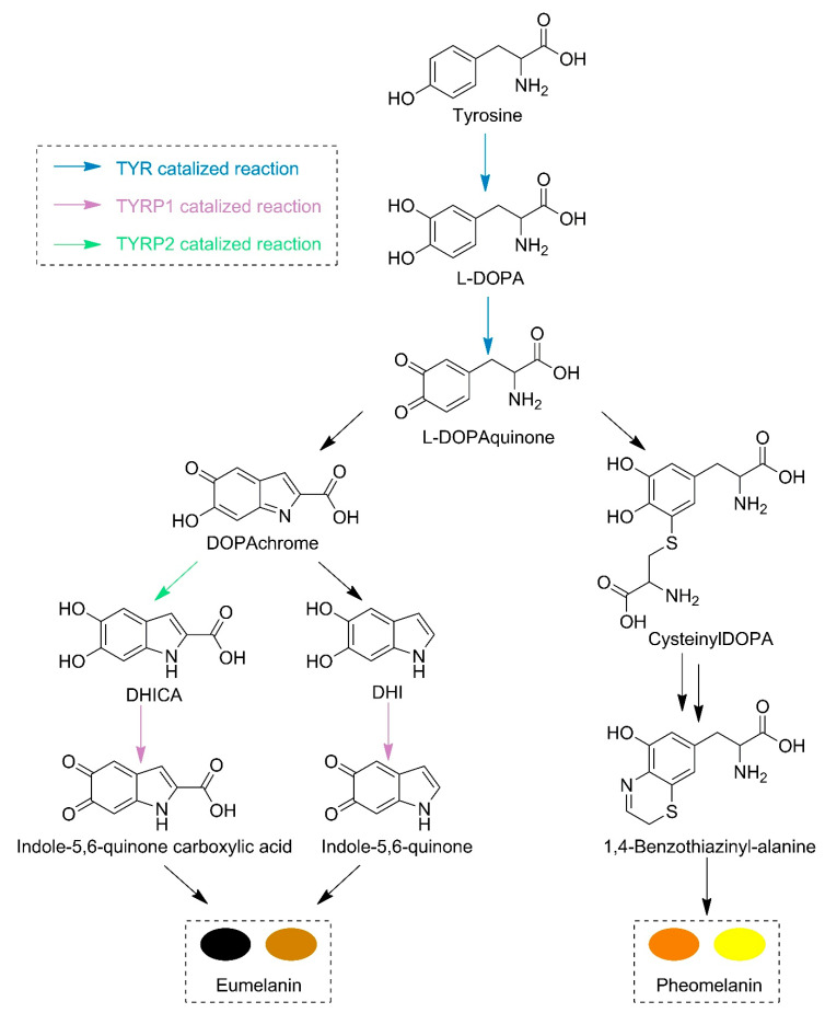
The three-dimensional structure of tyrosinase has been crystallized from many species but not from Homo sapiens. Tyrosinase is a key enzyme in melanin biosynthesis, being an important target for melanoma and skin-whitening cosmetics. Several studies employed the structure of tyrosinase from Agaricus bisporus as a model enzyme. Recently, 98% of human genome proteins were elucidated by AlphaFold. Herein, the AlphaFold structure of human tyrosinase and the previous model were compared. Moreover, tyrosinase-related proteins 1 and 2 were included, along with inhibition studies employing kojic and cinnamic acids. Peptides are widely studied for their inhibitory activity of skin-related enzymes. Cyanophycin is an amino acid polymer produced by cyanobacteria and is built of aspartic acid and arginine; arginine can be also replaced by other amino acids. A new set of cyanophycin-derived dipeptides was evaluated as potential inhibitors. Aspartate-glutamate showed the strongest interaction and was chosen as a leading compound for future studies.
Keywords: AlphaFold; molecular docking; tyrosinase; tyrosinase-related protein 1; tyrosinase-related protein 2.
Conflict of interest statement
The authors declare no conflict of interest. The funders had no role in the design of the study; in the collection, analyses, or interpretation of data; in the writing of the manuscript; or in the decision to publish the results.
Figures
Similar articles
-
Discovery of Potent Cysteine-Containing Dipeptide Inhibitors against Tyrosinase: A Comprehensive Investigation of 20 × 20 Dipeptides in Inhibiting Dopachrome Formation.J Agric Food Chem. 2015 Jul 15;63(27):6181-8. doi: 10.1021/acs.jafc.5b01026. Epub 2015 Jun 26. J Agric Food Chem. 2015. PMID: 26083974
-
Tyrosinase inhibition and anti-melanin generation effect of cinnamamide analogues.Bioorg Chem. 2019 Jun;87:43-55. doi: 10.1016/j.bioorg.2019.03.001. Epub 2019 Mar 4. Bioorg Chem. 2019. PMID: 30856375
-
Antioxidant, anti-tyrosinase and anti-melanogenic effects of (E)-2,3-diphenylacrylic acid derivatives.Bioorg Med Chem. 2019 Jun 1;27(11):2192-2200. doi: 10.1016/j.bmc.2019.04.020. Epub 2019 Apr 15. Bioorg Med Chem. 2019. PMID: 31027707
-
Naturally occurring and synthetic peptides: Efficient tyrosinase inhibitors.J Pept Sci. 2021 Jul;27(7):e3329. doi: 10.1002/psc.3329. Epub 2021 Apr 15. J Pept Sci. 2021. PMID: 33860571 Review.
-
Biomedical applications of tyrosinases and tyrosinase inhibitors.Enzymes. 2024;56:261-280. doi: 10.1016/bs.enz.2024.05.005. Epub 2024 Sep 7. Enzymes. 2024. PMID: 39304289 Review.
Cited by
-
Human Tyrosinase Displayed on the Surface of Chinese Hamster Ovary Cells for Ligand Fishing of Tyrosinase Inhibitors from Medicinal Plants.Molecules. 2024 Dec 25;30(1):30. doi: 10.3390/molecules30010030. Molecules. 2024. PMID: 39795088 Free PMC article.
References
-
- Solano F. Melanins: Skin Pigments and Much More—Types, Structural Models, Biological Functions, and Formation Routes. New J. Sci. 2014;2014:1–28. doi: 10.1155/2014/498276. - DOI
-
- Lapedriza A., Petratou K., Kelsh R.N. Neural Crest Cells and Pigmentation. Neural Crest Cells Evol. Dev. Dis. 2014;Chapter 14:287–311. doi: 10.1016/B978-0-12-401730-6.00015-6. - DOI
MeSH terms
Substances
Grants and funding
LinkOut - more resources
Full Text Sources
Other Literature Sources
Research Materials
