Review
. 2022 Mar 19;23(6):3338.
doi: 10.3390/ijms23063338.

Coagulopathy and Fibrinolytic Pathophysiology in COVID-19 and SARS-CoV-2 Vaccination

Affiliations
Review

Coagulopathy and Fibrinolytic Pathophysiology in COVID-19 and SARS-CoV-2 Vaccination

Shinya Yamada et al. Int J Mol Sci. .

Abstract

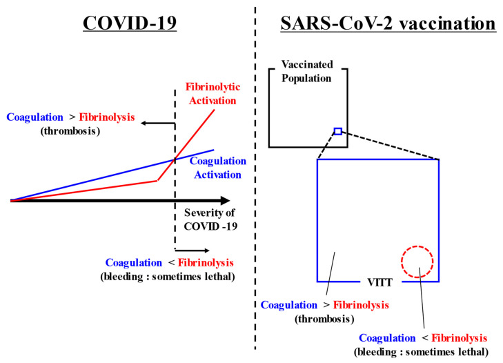
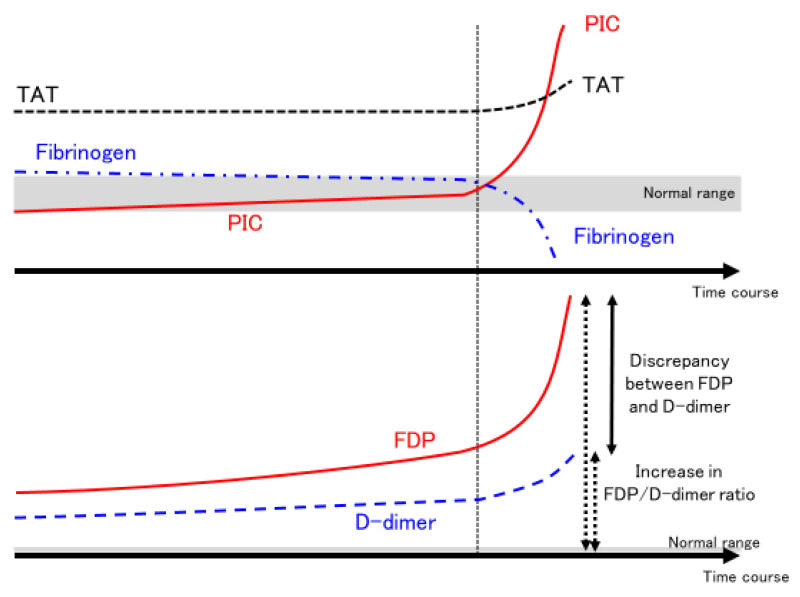
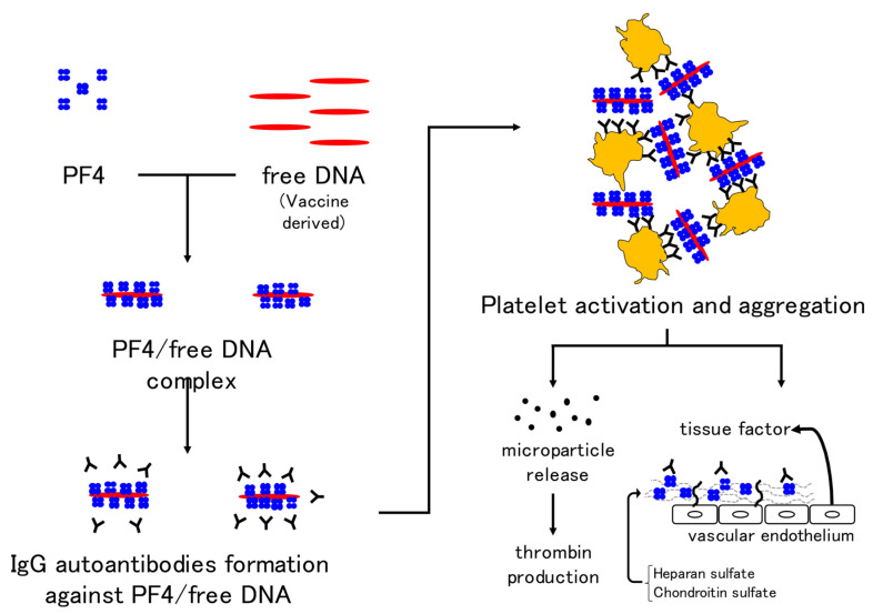
Coronavirus Disease 2019 (COVID-19) is caused by severe acute respiratory syndrome coronavirus 2 (SARS-CoV-2) and is frequently complicated by thrombosis. In some cases of severe COVID-19, fibrinolysis may be markedly enhanced within a few days, resulting in fatal bleeding. In the treatment of COVID-19, attention should be paid to both coagulation activation and fibrinolytic activation. Various thromboses are known to occur after vaccination with SARS-CoV-2 vaccines. Vaccine-induced immune thrombotic thrombocytopenia (VITT) can occur after adenovirus-vectored vaccination, and is characterized by the detection of anti-platelet factor 4 antibodies by enzyme-linked immunosorbent assay and thrombosis in unusual locations such as cerebral venous sinuses and visceral veins. Treatment comprises high-dose immunoglobulin, argatroban, and fondaparinux. Some VITT cases show marked decreases in fibrinogen and platelets and marked increases in D-dimer, suggesting the presence of enhanced-fibrinolytic-type disseminated intravascular coagulation with a high risk of bleeding. In the treatment of VITT, evaluation of both coagulation activation and fibrinolytic activation is important, adjusting treatments accordingly to improve outcomes.

Keywords: COVID-19; SARS-CoV-2 vaccine; coagulopathy; enhanced-fibrinolytic-type DIC; fibrinolysis; nafamostat.


Conflict of interest statement

The authors declare no conflict of interest.

Figures

Figure 1
Figure 1
Summarizing figure. COVID-19 causes coagulation activation depending on its severity. In addition, some cases of severe COVID-19 have markedly increased fibrinolysis. Thrombosis appears as the main symptom when coagulation activation exceeds fibrinolytic activation. Conversely, bleeding appears as the main symptom when fibrinolytic activation exceeds coagulation activation. After SARS-CoV-2 vaccination, VITT rarely occurs. In most cases, coagulation activation exceeds fibrinolytic activation and thrombosis occurs, but in some cases, bleeding appears when fibrinolytic activation exceeds coagulation activation. Abbreviations: COVID-19, coronavirus disease 2019; SARS-CoV-2, severe acute respiratory syndrome coronavirus 2; VITT, vaccine-induced immune thrombotic thrombocytopenia.
Figure 2
Figure 2
Dynamic changes in fibrinolytic pathophysiology in some severe COVID-19 cases. The time course of coagulation markers in COVID-19 patients with sudden enhancement of fibrinolytic activation and severe bleeding are shown. In some severe COVID-19 patients, PIC suddenly increases (PIC surge) at a certain point (around 7–10 days after admission to the ICU, as indicated by the vertical dotted line). At the same time, fibrinogen decreases significantly, but the degree of increase in TAT does not change significantly (upper panel). Meanwhile, FDP increases significantly, while D-dimer increases only mildly. Both solid and dotted arrows indicate the FDP/D-dimer ratio (lower panel). As PIC increases and fibrinogen decreases, discrepancy between FDP and D-dimer or the FDP/D-dimer ratio increases. These changes in coagulation markers are suggestive of enhanced-fibrinolytic-type DIC. Abbreviations: COVID-19, coronavirus disease 2019; ICU, intensive care unit; PIC, plasmin–α2 plasmin inhibitor complex; TAT, thrombin–antithrombin complex; FDP, fibrin/fibrinogen degradation products; DIC, disseminated intravascular coagulation.
Figure 3
Figure 3
Mechanism of VITT development in adenovirus vector vaccines. Immunoglobulin G autoantibodies against the complex of platelet factor 4 and free DNA in the vaccine result in platelet activation and aggregation. In addition, the release of microparticles and activation of coagulation by vascular endothelial cells are thought to be mechanisms underlying thrombus formation in VITT. Abbreviations: VITT, vaccine-induced immune thrombotic thrombocytopenia; PF4, platelet factor 4.
Figure 4
Figure 4
Relationship between VITT and fibrinolytic pathophysiology. We plotted fibrinogen on the horizontal axis and D-dimer on the vertical axis for 30 definitive VITT cases (using data from Reference [222]) in which both fibrinogen and D-dimer values were known. The lower the level of fibrinogen, the higher the level of D-dimer. In particular, patients with blue dotted squares showed a decrease in fibrinogen and a marked increase in D-dimer, suggesting the complication of enhanced-fibrinolytic-type DIC (a subject for further investigation). If anticoagulants are administered to the same intensity as in other cases, the risk of bleeding is considered high. To confirm the diagnosis of enhanced-fibrinolytic-type DIC, measurement of not only the coagulation activation marker TAT, but also the fibrinolysis activation marker PIC is essential. Patients with markedly decreased α2PI are at high risk of bleeding. Importantly, in enhanced-fibrinolytic-type DIC, the increase in FDP is more prominent than the increase in D-dimer (discrepancy between FDP and D-dimer). Antithrombin, as a coagulation inhibitor, does not often decrease, even in patients with a marked decrease in α2PI, a fibrinolytic inhibitor (except in patients with reduced hepatic reserve). Abbreviations: VITT, vaccine-induced immune thrombotic thrombocytopenia; DIC, disseminated intravascular coagulation; TAT, thrombin–antithrombin complex; PIC, plasmin–α2 plasmin inhibitor complex; α2 PI, α2 plasmin inhibitor; FDP, fibrin/fibrinogen degradation products.

Similar articles

Cited by

References

    1. Geerts W., Selby R. Prevention of Venous Thromboembolism in the ICU. Chest. 2003;124:357S–363S. doi: 10.1378/chest.124.6_suppl.357S. - DOI - PubMed
    1. Horiuchi H., Morishita E., Urano T., Yokoyama K. The Questionnaire-survey Joint Team on The COVID-19-related thrombosis COVID-19-Related Thrombosis in Japan: Final Report of a Questionnaire-Based Survey in 2020. J. Atheroscler. Thromb. 2021;28:406–416. doi: 10.5551/jat.RPT001. - DOI - PMC - PubMed
    1. Kerbikov O., Orekhov P., Borskaya E., Nosenko N. High incidence of venous thrombosis in patients with moderate-to-severe COVID-19. Int. J. Hematol. 2021;113:344–347. doi: 10.1007/s12185-020-03061-y. - DOI - PMC - PubMed
    1. Hunter P.R. Thrombosis after COVID-19 vaccination. BMJ. 2021;373:n958. doi: 10.1136/bmj.n958. - DOI - PubMed
    1. Nicosia R.F., Ligresti G., Caporarello N., Akilesh S., Ribatti D. COVID-19 Vasculopathy: Mounting Evidence for an Indirect Mechanism of Endothelial Injury. Am. J. Pathol. 2021;191:1374–1384. doi: 10.1016/j.ajpath.2021.05.007. - DOI - PMC - PubMed

MeSH terms