. 2022 Mar 21;23(6):3373.
doi: 10.3390/ijms23063373.

Two Male-Specific Antimicrobial Peptides SCY2 and Scyreprocin as Crucial Molecules Participated in the Sperm Acrosome Reaction of Mud Crab Scylla paramamosain

Affiliations

Two Male-Specific Antimicrobial Peptides SCY2 and Scyreprocin as Crucial Molecules Participated in the Sperm Acrosome Reaction of Mud Crab Scylla paramamosain

Ying Yang et al. Int J Mol Sci. .

Abstract

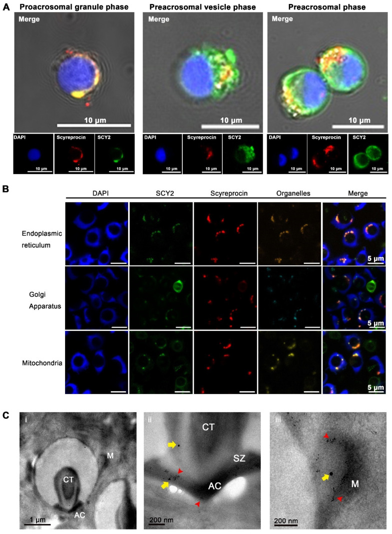
Antimicrobial peptides (AMPs) identified in the reproductive system of animals have been widely studied for their antimicrobial activity, but only a few studies have focused on their physiological roles. Our previous studies have revealed the in vitro antimicrobial activity of two male gonadal AMPs, SCY2 and scyreprocin, from mud crab Scylla paramamosain. Their physiological functions, however, remain a mystery. In this study, the two AMPs were found co-localized on the sperm apical cap. Meanwhile, progesterone was confirmed to induce acrosome reaction (AR) of mud crab sperm in vitro, which intrigued us to explore the roles of the AMPs and progesterone in AR. Results showed that the specific antibody blockade of scyreprocin inhibited the progesterone-induced AR without affecting intracellular Ca2+ homeostasis, while the blockade of SCY2 hindered the influx of Ca2+. We further showed that SCY2 could directly bind to Ca2+. Moreover, progesterone failed to induce AR when either scyreprocin or SCY2 function was deprived. Taken together, scyreprocin and SCY2 played a dual role in reproductive immunity and sperm AR. To our knowledge, this is the first report on the direct involvement of AMPs in sperm AR, which would expand the current understanding of the roles of AMPs in reproduction.

Keywords: SCY2; acrosome reaction; antimicrobial peptide (AMP); fertilization; invertebrate; progesterone; scyreprocin; sperm.


Conflict of interest statement

The authors declare no conflict of interest.

Figures

Figure 1
Figure 1
Scyreprocin and SCY2 expressed in reproductive system of adult male mud crabs and transferred to female spermathecae via mating. (A) Scyreprocin transcriptional expression level in adult male (n = 3) and female (n = 3) Scylla paramamosain under natural conditions. Data are presented as the mean ± standard deviation (SD). * p < 0.05, one-way analysis of variance (ANOVA) and Tukey post-test. (B) Scyreprocin expression profiles in different tissues of adult male and female crabs (n = 3). (C) Scyreprocin expression profiles in semen (sperm and seminal plasma) collected from adult and juvenile males (n = 3). BW, body weight. (D) In situ expression of SCY2 (green) and scyreprocin (red) in testes of juvenile and adult males. (E) In situ expression of SCY2 (green) and scyreprocin (red) in spermathecae of pre- and post-mating females. In panels (D,E), nucleus is shown in blue color. Abbreviations: Br, brain; Gi, gill; St, stomach; Mg, midgut; Ht, heart; Hp, hepatopancreas; SE, subcuticular epidermis; Ne, thoracic ganglion mass; Mu, muscle; Es, eyestalk; Hc, hemolymph cell; T, testis; AVD, anterior vas deferens; SV, seminal vesicle; PVD, posterior vas deferens; ED, ejaculatory duct; PED, posterior ejaculatory duct; P, penis; S, spermatheca; OA, ovary; Vg, vagina.
Figure 2
Figure 2
Scyreprocin and SCY2 responded to bacterial infections. (A) Morphological changes induced by recombinant scyreprocin (rScyreprocin) and SCY2 (rSCY2) in Pseudomonas putida isolate X1 (n = 3). P. putida isolate X1 (5 × 10⁵ cfu mL−1) was incubated with rScyreprocin (2 μM) or rSCY2 (4 μM) for 30 min and observed by a scanning electron microscopy. (B) Induction of SCY2 and scyreprocin expression levels in in vitro cultured spermatophores after P. putida isolate X1 challenge (n = 3). The in vitro cultured spermatophore were incubated with P. putida isolate X1 (100 cfu well−1) for 24 h before subjected to immunofluorescence assay. (C) Induction of SCY2 and scyreprocin expression levels in testis (T) and ejaculatory duct (ED) by in vivo P. putida isolate X1 challenge (n = 3). Adult male crabs were challenged with P. putida isolate X1 (3 × 10³ cfu crab−1). After 24 h, T and ED were sampled and subjected to Western blot analysis. (D) Quantification of the blots in (C) by ImageJ. Data are presented as the mean ± standard deviation (SD). * p < 0.05, two-way analysis of variance (ANOVA) and Bonferroni post-test.
Figure 3
Figure 3
Subcellular location of scyreprocin and SCY2 in male gametes. (A) In situ expression of scyreprocin (red) and SCY2 (green) in spermatids at different spermiogenesis stages, nucleus was stained with DAPI (blue). In vitro cultured testicular cells (seeded at 2 × 10⁶ cells well−1 for 3 days) were subjected to immunofluorescence assay. (B) SCY2 and scyreprocin co-localized with organelles in sperm. Sperm were freshly isolated from seminal vesicles of adult male crabs and subjected to immunofluorescence assay. (C) In situ expression of scyreprocin and SCY2 observed by transmission electron microscope (TEM) in mud crab sperm: i, intact sperm; ii, apical cap (AC); iii, mitochondria (M). Red arrows: scyreprocin; yellow arrows, SCY2. Abbreviations: SZ, sub-cap zone; CT, central tube.
Figure 4
Figure 4
Progesterone (PG) was a crucial acrosome reaction (AR)-induced substance for crab sperm. (A) Schematic presentation of the AR ratio (%AR) evaluation on the sperm collected from male and female crabs. N, spermathecae; ASW, artificial seawater; Ca2+-FASW, Ca2+-free ASW. (B) Statistical analysis on %AR of sperm collected from female spermathecae and male gonads (n = 3). Data are presented as the mean ± standard deviation (SD). * p < 0.05, one-way analysis of variance (ANOVA) and Tukey post-test; n.s., not significant. M, male; F, female. (C) Flow cytometry assessment on %AR of sperm collected from female spermathecae and male gonads (n = 3). Sperm samples were treated with ASW, Ca2+-FASW (male- and female-derived sperm), or ASW containing 20 μg mL−1 PG (male-derived sperm) for 24 h before subjected to flow cytometry analysis. (D) Ultrastructural changes of crab sperm during AR observed by scanning electron microscope (SEM). Male-derived sperm after PG treatment in (C) were subjected for SEM observation: 1–2, unreacted sperm; 3, acrosome protruding stage; 4, acrosomal vesicle valgus stage; 5, central tube extension stage; 6, reacted sperm. (E) Changes in PG level in spermatheca (S) and ovary (OA) at pre- and post-mating stages. Spermathecae and ovaries from un-mated females, female crabs at the day after mating, post-mating stage I, II, III, pre-ovulation, and post-ovulation stage, were collected (n = 6). The samples (~30 mg) were subjected to PG level analysis. Data are presented as the mean ± SD.
Figure 5
Figure 5
Expression pattern of scyreprocin and SCY2 in sperm during the acrosome reaction (AR). (A) Subcellular localization of scyreprocin and SCY2 in sperm at different AR stages (blue, nucleus; red, scyreprocin; green, SCY2). Male-derived sperm were treated with artificial seawater (ASW) containing 20 μg mL−1 PG for 24 h before subjected to immunofluorescence assay. (B) Subcellular localization of scyreprocin (red arrows) in sperm at different AR stages, from transmission electron microscopy (TEM) observation. Dashed lines indicate the zoom-in regions. Abbreviations: AC, apical cap; CT, central tube; M, mitochondria; SZ, sub-cap zone; AV, acrosomal vesicle; N, nucleus; AVM, acrosomal vesicle membrane.
Figure 6
Figure 6
Scyreprocin and SCY2 functioned as critical molecules in progesterone (PG)-induced sperm acrosome reaction (AR). (A) Schematic presentation of the AR ratio (%AR) and intracellular Ca2+ concentration ({Ca2+}i) evaluation of the sperm collected from male and female crabs. (B) Flow cytometry analysis of the sperm %AR after different treatments. Male-derived sperm samples (~1 × 10⁶ cells mL−1) were pre-treated with SCY2 antibody (1:500) and/or scyreprocin antibody (1:1000) for 2 h and incubated with PG (50 μg mL−1 in artificial seawater) for 22 h. Samples were subjected to flow cytometry analysis. (C) Statistical analyses of the flow cytometry data presented in (B) (n = 3). Data are presented as the mean ± standard deviation (SD). (D) Evaluation of {Ca2+}i in sperm samples in (B) (n = 3). In panels (C,D), “+” represents the addition of the corresponding component, data are presented as the mean ± SD. Letters denote significant differences, one-way analysis of variance (ANOVA), and Tukey post-test.
Figure 7
Figure 7
Interplay of scyreprocin, SCY2, progesterone (PG), and Ca2+. (A) Comparative examination of PG binding capacity of rScyreprocin, rSCY2 and rScyreprocin/rSCY2 mixture by a modified enzyme-linked immunosorbent assay (ELISA) (n = 3). (B) Schematic presentation of the intracellular Ca2+ concentration ({Ca2+}i) evaluation and acrosome reaction ratio (%AR) analysis. (C) Evaluation of {Ca2+}i in the sperm pretreated with SCY2 antibody or Ni2+ (n = 3). Sperm (2 × 10⁷ sperm mL−1) were pre-treated with Ni2+ (5 μM) or SCY2 antibody (1:500) for 2 h, and incubated with PG (50 μg mL−1 in artificial seawater) for 22 h. Samples were subjected to {Ca2+}i evaluation. (D) Flow cytometry analysis of the %AR (n = 3) of the samples in (C). (E) Statistical analysis of the data presented in (D). In panels C and E, data are presented as the mean ± SD. Letters denote significant differences, one-way ANOVA and Tukey post-test. (F) Electrophoretic mobility shift assays on the binding properties of rSCY2 with Ca2+, Mg2+, and Ni2+. rSCY2 (2 μg) was incubated in Tris-HCl containing CaCl2, MgCl2, or NiCl2 (0.1 mM) for 3 h and then supplemented with EGTA or EDTA (0.1 mM) for 10 min. Samples were subjected to native gel electrophoresis.
Figure 8
Figure 8
Roles of scyreprocin and SCY2 in the sperm acrosome reaction (AR) of Scylla paramamosain. Adult male crabs expressed scyreprocin and SCY2 in semen, which were then transferred to female spermatheca via mating. Scyreprocin and SCY2 in seminal plasma were proved to maintain gamete health by exerting antimicrobial activity. In sperm, scyreprocin and SCY2 showed co-localization on the apical cap and mitochondria, and are proven to participate in the initiation of progesterone-induced AR. In un-reacted sperm, SCY2 was responsible for maintaining intracellular Ca2+ homeostasis. Upon sperm–egg attachment, scyreprocin bound to progesterone, with SCY2 cooperatively strengthening the binding affinity. SCY2 bound to extracellular Ca2+ and transported it into the sperm. The increase in {Ca2+}i ultimately initiated AR and allowed completion of sperm–egg fusion. Abbreviations: AC, apical cap; AV, acrosomal vesicle; {Ca2+}i, intracellular Ca2+ concentration.
Figure 9
Figure 9
Brief comparison of sperm acrosome reaction (AR) mechanism in mammals, sea urchin, and mud crab. In mammals, progesterone and egg zona pellucida proteins (ZPs) interact with their corresponding receptors (i.e., CatSper, ZP receptors) on the sperm membrane, activate CatSper, and induce Ca2+ influx. Increase in intracellular Ca2+ concentration ({Ca2+}i) then leads to the release of Ca2+ from intracellular Ca2+ store, and thus completes sperm AR. In sea urchins, sperm AR is induced by the interaction of fucose sulfated glycoconjugate from egg-coat (FSG) and its specific receptor (REJ) on the sperm membrane, which opens a Ca2+-selective channel and a store-operated Ca2+ channel and leads to vesicular fusion. In mud crab, progesterone interacts with scyreprocin on the sperm surface, thus inducing Ca2+ influx mediated by SCY2 and initiating sperm AR. The AR molecular basis of mud crab Scylla paramamosain revealed in the present study is different from that of other species.

Similar articles

Cited by

References

    1. Stival C., Puga Molina L.D.C., Paudel B., Buffone M., Visconti P., Krapf D. Sperm capacitation and acrosome reaction in mammalian sperm. Sperm Acrosome Biog. Funct. Dur. Fertil. 2016;220:93–106. - PubMed
    1. Dan J.C. Studies on the acrosome. I. Reaction to egg-water and other stimuli. Biol. Bull. 1952;103:54–66. doi: 10.2307/1538405. - DOI
    1. Ryuzo Y., Gary C., Takahiro M., Tadashi A., Tatsuo H., Carol V., Murali P., Frederick G., Hajime M., Tina W. Sperm attractant in the micropyle region of fish and insect eggs. Biol. Reprod. 2013;88:47. doi: 10.1095/biolreprod.112.105072. - DOI - PubMed
    1. Wilson K.L., Fitch K.R., Bafus B.T., Wakimoto B.T. Sperm plasma membrane breakdown during Drosophila fertilization requires sneaky, an acrosomal membrane protein. Development. 2006;133:4871–4879. doi: 10.1242/dev.02671. - DOI - PubMed
    1. Hirohashi N., Yanagimachi R. Sperm acrosome reaction: Its site and role in fertilization. Biol. Reprod. 2018;99:127–133. doi: 10.1093/biolre/ioy045. - DOI - PubMed