. 2022 Mar 21;23(6):3388.
doi: 10.3390/ijms23063388.

Swelling, Protein Adsorption, and Biocompatibility In Vitro of Gel Beads Prepared from Pectin of Hogweed Heracleum sosnówskyi Manden in Comparison with Gel Beads from Apple Pectin

Affiliations

Swelling, Protein Adsorption, and Biocompatibility In Vitro of Gel Beads Prepared from Pectin of Hogweed Heracleum sosnówskyi Manden in Comparison with Gel Beads from Apple Pectin

Sergey Popov et al. Int J Mol Sci. .

Abstract

The study aims to develop gel beads with improved functional properties and biocompatibility from hogweed (HS) pectin. HS4 and AP4 gel beads were prepared from the HS pectin and apple pectin (AP) using gelling with calcium ions. HS4 and AP4 gel beads swelled in PBS in dependence on pH. The swelling degree of HS4 and AP4 gel beads was 191 and 136%, respectively, in PBS at pH 7.4. The hardness of HS4 and AP4 gel beads reduced 8.2 and 60 times, respectively, compared with the initial value after 24 h incubation. Both pectin gel beads swelled less in Hanks' solution than in PBS and swelled less in Hanks' solution containing peritoneal macrophages than in cell-free Hanks' solution. Serum protein adsorption by HS4 and AP4 gel beads was 118 ± 44 and 196 ± 68 μg/cm2 after 24 h of incubation. Both pectin gel beads demonstrated low rates of hemolysis and complement activation. However, HS4 gel beads inhibited the LPS-stimulated secretion of TNF-α and the expression of TLR4 and NF-κB by macrophages, whereas AP4 gel beads stimulated the inflammatory response of macrophages. HS4 gel beads adsorbed 1.3 times more LPS and adhered to 1.6 times more macrophages than AP4 gel beads. Thus, HS pectin gel has advantages over AP gel concerning swelling behavior, protein adsorption, and biocompatibility.

Keywords: TNF-α; apple pectin; complement activation; gel beads; hemolysis; hogweed pectin; peritoneal macrophages; protein adsorption; swelling.


Conflict of interest statement

The authors declare no conflict of interest.

Figures

Figure 1
Figure 1
Photographs of wet AP4 (A), HS4 (C,) and dried AP4 (B), HS4 (D) gel beads.
Figure 2
Figure 2
Mean (n = 15) force overtime curve of the wet AP4 and HS4 gel beads.
Figure 3
Figure 3
Swelling degree of AP4 (A) and HS4 (B) gel beads incubating in PBS of different pH for 24 h. The data are presented as the mean (n = 10). a p < 0.05 vs. PBS pH 7.4; b p < 0.05 vs. the corresponding AP4 gel beads.
Figure 4
Figure 4
Variation in the pH values during incubation of AP4 (A) and HS4 (B) gel beads in PBS at different pH for 24 h. The data are presented as the mean ± S.D. (n = 5). a p < 0.05 vs. corresponding initial pH; b p < 0.05 vs. the corresponding AP4 gel beads levels.
Figure 5
Figure 5
Swelling degree of AP4 (A) and HS4 (B) gel beads incubating in PBS at pH 7.4 and Hanks’ solution (pH 7.4) for 24 h. The data are presented as the mean (n = 10). a p < 0.05 vs. PBS pH 7.4; b p < 0.05 vs. the corresponding AP4 gel beads.
Figure 6
Figure 6
Mean (n = 15) force overtime curve of the AP4 (A) and HS4 (B) gel beads incubated in Hanks’ solution for 24 h.
Figure 7
Figure 7
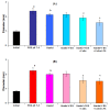
Effect of composition of incubating medium on the swelling of AP4 (A) and HS4 (B) gel beads. Dried gel beads (“Initial”) were incubated for 4 h in PBS (“PBS” pH 7.4); Hanks’ solution at pH 7.4 (“Hanks’”); Hanks’ solution supplemented with 10% fetal bovine serum (“Hanks’+FBS”); Hanks’ solution supplemented with 10% fetal bovine serum and 2 × 10⁶ cells/mL peritoneal macrophages (“Hanks’+FBS+Cells”); Hanks’ solution supplemented with 10% fetal bovine serum, 2 × 10⁶ peritoneal macrophages, and 10 μg/mL of lipopolysaccharide (“Hanks’+FBS+Cells+LPS”). The data are presented as the mean ± S.D. (n = 12). a p < 0.05 vs. “Initial”; b p < 0.05 vs. PBS pH 7.4; c p < 0.05 vs. Hanks’+FBS.
Figure 8
Figure 8
Mean (n = 15) force overtime curve of the AP4 (A) and HS4 (B) gel beads incubated in PBS (“PBS pH 7.4”); Hanks’ solution at pH 7.4 (“Hanks’”); Hanks’ solution supplemented with 10% FBS (“Hanks’+FBS”); Hanks’ solution supplemented with 10% FBS and 2 × 10⁶ cells/mL peritoneal macrophages (“Hanks’+FBS+Mφ”); Hanks’ solution supplemented with 10% fetal bovine serum, 2 × 10⁶ peritoneal macrophages, and 10 μg/mL of LPS (“Hanks’+FBS+Mφ+LPS”).
Figure 9
Figure 9
The BSA adsorption by AP4 (A) and HS4 (B) gel beads incubating in PBS at different pH for 24 h. The data are presented as the mean ± S.D. (n = 5). a p < 0.05 vs. PBS at pH 7.4; b p < 0.05 vs. the corresponding AP4 gel beads. The BSA adsorption is expressed as the relative protein concentration Ct/C0, where C0 and Ct are the initial and remaining concentrations of BSA in PBS at different time points after incubation, respectively.
Figure 10
Figure 10
The BSA adsorption expressed per unit surface area by pectin gel beads incubating in PBS at different pH for 24 h. The data are presented as the mean ± S.D. (n = 5). a p < 0.05 vs. PBS pH 7.4; b p < 0.05 vs. the corresponding AP4 gel beads.
Figure 11
Figure 11
Variation in the pH values during incubation of AP4 and HS4 gel beads for 24 h in acetate buffer solutions of pH 3.7 (A) and 5.0 (B). The data are presented as the mean ± S.D. (n = 5).
Figure 12
Figure 12
The BSA adsorption by AP4 and HS4 gel beads incubating for 24 h in acetate buffer solutions of pH 3.7 (A) and 5.0 (B). The data are presented as the mean ± S.D. (n = 5). a p < 0.05 vs. initial time point; b p < 0.05 vs. the corresponding AP4 gel beads. The BSA adsorption is expressed as the relative protein concentration Ct/C0, where C0 and Ct are the initial and remaining concentrations of BSA in PBS at different time points after incubation, respectively.
Figure 13
Figure 13
The serum proteins adsorption by AP4 and HS4 gel beads incubating for 24 h in Hanks’ solution supplemented with 10% FBS. The data are presented as the mean ± S.D. a p < 0.05 vs. the corresponding AP4 gel beads. The protein adsorption is expressed as the relative protein concentration Ct/C0, where C0 and Ct are the initial and remaining concentrations in Hanks’ solution at different time points after incubation, respectively.
Figure 14
Figure 14
The serum proteins adsorption expressed per unit surface area by AP4 and HS4 gel beads incubating for 4 (A) and 24 h (B) in Hanks’ solution supplemented with 10% FBS. The data are presented as the mean ± S.D. (n = 5). a p < 0.05 vs. the corresponding AP4 gel beads.
Figure 15
Figure 15
Effect of AP4 and HS4 gel beads on the C3a production in the whole blood in vitro. Results are presented as the mean ± S.D. (n = 8). a and bp < 0.05 vs. NaCl and Zymosan, respectively.
Figure 16
Figure 16
LPS adsorption (A) and macrophage adhesion (B) on the AP4 and HS4 gel beads during incubation for 4 h in Hanks’ solution supplemented with 10% FBS, 10 μg/mL LPS, and 2 × 10⁶ peritoneal macrophages. The data are presented as the mean ± S.D. (n = 12). a and bp < 0.05 vs. Control and AP4 gel beads, respectively. C4/C0—relative concentration, where C0 and C4 are the initial and remaining concentrations of LPS in Hanks’ solution after 4 h of incubation, respectively.
Figure 17
Figure 17
The impact of pectin gel beads on inflammatory proteins levels in mouse peritoneal macrophages. Bar graphs show TNF-α concentration (A) and TLR4 (C), and NF-κB (D) protein levels, which are normalized to the β-actin protein level. (B)—representative bands of NF-κB, TLR4, and β-actin. The results are expressed as the mean ± S.D. (n = 8). a and bp < 0.05 vs. Control and AP4 gel beads, respectively.

Similar articles

Cited by

References

    1. Moslemi M. Reviewing the recent advances in application of pectin for technical and health promotion purposes: From laboratory to market. Carbohydr. Polym. 2021;254:117324. doi: 10.1016/j.carbpol.2020.117324. - DOI - PubMed
    1. Cascone S., Lamberti G. Hydrogel-based commercial products for biomedical applications: A review. Int. J. Pharm. 2020;573:118803. doi: 10.1016/j.ijpharm.2019.118803. - DOI - PubMed
    1. Neves S.C., Moroni L., Barrias C.C., Granja P.L. Leveling up hydrogels: Hybrid systems in tissue engineering. Trends Biotechnol. 2019;38:292–315. doi: 10.1016/j.tibtech.2019.09.004. - DOI - PubMed
    1. Lara-Espinoza C., Carvajal-Millán E., Balandran-Quintana R., Lopez-Franco Y., Rascon-Chu A. Pectin and pectin-based composite materials: Beyond food texture. Molecules. 2018;23:942. doi: 10.3390/molecules23040942. - DOI - PMC - PubMed
    1. Minzanova S.T., Mironov V.F., Arkhipova D.M., Khabibullina A.V., Mironova L.G., Zakirova Y.M., Milyukov V.A. Biological activity and pharmacological application of pectic polysaccharides: A review. Polymers. 2018;10:1407. doi: 10.3390/polym10121407. - DOI - PMC - PubMed

LinkOut - more resources