Review
. 2022 Mar 11;19(6):3331.
doi: 10.3390/ijerph19063331.

The Role of 3D Printing in Planning Complex Medical Procedures and Training of Medical Professionals-Cross-Sectional Multispecialty Review

Affiliations
Review

The Role of 3D Printing in Planning Complex Medical Procedures and Training of Medical Professionals-Cross-Sectional Multispecialty Review

Jarosław Meyer-Szary et al. Int J Environ Res Public Health. .

Abstract

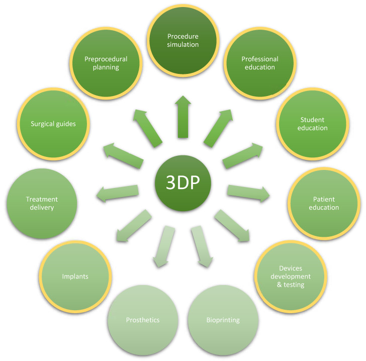
Medicine is a rapidly-evolving discipline, with progress picking up pace with each passing decade. This constant evolution results in the introduction of new tools and methods, which in turn occasionally leads to paradigm shifts across the affected medical fields. The following review attempts to showcase how 3D printing has begun to reshape and improve processes across various medical specialties and where it has the potential to make a significant impact. The current state-of-the-art, as well as real-life clinical applications of 3D printing, are reflected in the perspectives of specialists practicing in the selected disciplines, with a focus on pre-procedural planning, simulation (rehearsal) of non-routine procedures, and on medical education and training. A review of the latest multidisciplinary literature on the subject offers a general summary of the advances enabled by 3D printing. Numerous advantages and applications were found, such as gaining better insight into patient-specific anatomy, better pre-operative planning, mock simulated surgeries, simulation-based training and education, development of surgical guides and other tools, patient-specific implants, bioprinted organs or structures, and counseling of patients. It was evident that pre-procedural planning and rehearsing of unusual or difficult procedures and training of medical professionals in these procedures are extremely useful and transformative.

Keywords: 3D printing; additive manufacturing; cardiac surgery; cardiology; gynecology; head and neck surgery; mandible reconstruction; obstetrics; oncology; orthopedics; otolaryngology; outcomes; radiotherapy; simulation; surgery; three-dimensional printing; training; trauma; urology.


Conflict of interest statement

The authors declare no conflict of interest.

Figures

Figure 1
Figure 1
The number of PubMed indexed publications since 2000 (total 19,562) and fitting exponential trend line. Search query: 3D printing or additive manufacturing.
Figure 2
Figure 2
The steps in the process of 3D printing. (A). Source CT; (B). CT after segmentation (red); (C). Digital model (STL); (D). Print setup; (E). Raw print covered in support material; (F). Final 3D printed model. Detailed descriptions in text. (If not stated otherwise, images courtesy of Jarosław Meyer-Szary, Department of Pediatric Cardiology and Congenital Heart Defects, Faculty of Medicine, Medical University of Gdańsk, Gdańsk, Poland).
Figure 3
Figure 3
Examples of multi-part models. (A) a case of aortic ring, to visualize a compressed trachea by adjacent, abnormally positioned vessels, a cardiovascular model is printed in white and, separately segmented, trachea is printed green and fitted in place; (B) a case of neonate with a mediastinal tumor (white) compressing the cardiovascular structures (red), the superior vena cava (asterisk) and the right atrium (hashtag), the model was based on multimodality imaging owing to the fact that Angio CT has superior special and temporal resolution for cardiovascular structures while MRI has superior ability to differentiate soft tissue structures and tumors. Parts of the models coming from different modalities were assembled in post-production.
Figure 4
Figure 4
Breadth of medical applications of 3D printing. Highlighted in yellow are those in focus in this review. The list is non-exhaustive, and new applications are evolving.
Figure 5
Figure 5
Cardiovascular model of an interrupted aortic arch of a 3.5 kg neonate made to facilitate preoperative planning. SLS method was chosen for its superior handling of tiny details especially given no supports are necessary for the process. (A) Anterior view, dilated mammary arteries as part of collateral circulation (asterisk). (B) Posterior view, other collateral arteries (asterisk); top (curved) arrow—aortic arch; bottom (straight) arrow—distal part of a thoracic aorta; 2.5 cm gap between the arrows is where a surgeon needs to place anastomosis.
Figure 6
Figure 6
A set of two MRI-based models for planning and simulation of cardiovascular intervention—implantation of a Melody valve across a secondary pulmonary artery conduit stenosis (arrows) in an adult born with tetralogy of Fallot previously treated at the age of six by surgical implantation of pulmonary artery conduit, which has now become near-obstructed (arrows). (A) 3D-printed blood pool anatomical model including the pulmonary artery and aorta. (B) 3D-printed hollow model. (C) Fluoroscopic image of contrast-filled angioplasty balloon catheter across the stenosis. (D) Pre-dilatation fluoroscopic image of the implantation site (landing zone) using a similar balloon catheter during the actual intervention. The case was previously discussed in detail elsewhere [14].
Figure 7
Figure 7
Three-dimensional-printed model for transcatheter aortic valve implantation (TAVI) procedure training. (A) Parts for two different models at the post-processing stage—UV cured but waiting for support removal and assembly. (B) Assembled model with a catheter inserted. (C) Closeup of the left ventricle showing a balloon catheter inside. (D) View from the inside with the balloon catheter inflated. FA—femoral arteries, AbdA—abdominal aorta, ThA—thoracic aorta, AA—aortic arch, LV—left ventricle, BC—balloon catheter.
Figure 8
Figure 8
Three-dimensional printing in free fibula flap mandible reconstruction following odontogenic tumor resection: (A) Computer (CAD) plan of a reconstructed mandible; (B) Computer model and 3D print of fibula with planned construct segments marked out; (C) 3D-printed model next to actual patient’s fibula in vivo; (D) 3D-printed jig with osteotomy (cutting) guides on fibula; (E) segmented fibula in vivo and computer model with corresponding segments labeled; (F) assembled construct fixed with pre-bent metal reconstruction plate, prior to division of vascular pedicle; (G) CT image of patient three months following surgery. (Images courtesy of Dr. Chew Khong-Yik, Department of Plastic Surgery, Singapore General Hospital, Singapore).
Figure 9
Figure 9
Three-dimensional-printed models of brain aneurisms (arrows) based on digital subtraction angiography: (A) An Anterior Communicating Artery / Right Anterior Cerebral Artery A2 (AcomA/ACA A2 dex) aneurysm. (B) A giant aneurysm of the right internal carotid artery (rICA). rPCA – right Posterior Cerebral Artery, rMCA – right Middle Cerebral Artery.
Figure 10
Figure 10
Three-dimensional prints of kidney anatomy; (A) Virtual 3D reconstruction based on contrast-CT scan of the kidney (short arrow) with the tumor (long arrow). (B) Contrast-CT scan of the kidney (short arrow) with the tumor (long arrow). (C) Top—3D-printed model of the kidney (short arrow) with the tumor printed with a separate color (long arrow); Bottom—resected kidney (short arrow) with the tumor (long arrow). Nephrectomy was necessary due to the tumor penetrating into the renal calyx. The 3D print model corresponded to the actual size of the kidney and the tumor.
Figure 11
Figure 11
Applications of 3DP in radiotherapy. (A) An individually created hand bolus for total body irradiation before an allogenic stem cell transplant. (B) Boluses of different thicknesses (0.5 and 1 cm) with different infill percentages (60% and 20%) changing their physical properties.

References

    1. Antreas K., Piromalis D. Employing a Low-Cost Desktop 3D Printer: Challenges, and How to Overcome Them by Tuning Key Process Parameters. Int. J. Mech. Appl. 2021;10:11–19. doi: 10.5923/j.mechanics.20211001.02. - DOI
    1. Luo H., Meyer-Szary J., Wang Z., Sabiniewicz R., Liu Y. Three-Dimensional Printing in Cardiology: Current Applications and Future Challenges. Cardiol. J. 2017;24:436–444. doi: 10.5603/CJ.a2017.0056. - DOI - PubMed
    1. Garg R., Zahn E.M. Utility of Three-Dimensional (3D) Modeling for Planning Structural Heart Interventions (with an Emphasis on Valvular Heart Disease) Curr. Cardiol. Rep. 2020;22:125. doi: 10.1007/s11886-020-01354-6. - DOI - PubMed
    1. Meyer-Szary J., Woźniak-Mielczarek L., Sabiniewicz D., Sabiniewicz R. Feasibility of In-House Rapid Prototyping of Cardiovascular Three-Dimensional Models for Planning and Training Non-Standard Interventional Procedures. Cardiol. J. 2020;26:790–792. doi: 10.5603/CJ.2019.0115. - DOI - PMC - PubMed
    1. Scanlan A.B., Nguyen A.V., Ilina A., Lasso A., Cripe L., Jegatheeswaran A., Silvestro E., McGowan F.X., Mascio C.E., Fuller S., et al. Comparison of 3D Echocardiogram-Derived 3D Printed Valve Models to Molded Models for Simulated Repair of Pediatric Atrioventricular Valves. Pediatr. Cardiol. 2018;39:538–547. doi: 10.1007/s00246-017-1785-4. - DOI - PMC - PubMed

LinkOut - more resources