. 2022 Mar 9;15(6):2014.
doi: 10.3390/ma15062014.

Accelerated Endothelialization of Nanofibrous Scaffolds for Biomimetic Cardiovascular Implants

Affiliations

Accelerated Endothelialization of Nanofibrous Scaffolds for Biomimetic Cardiovascular Implants

Claudia Matschegewski et al. Materials (Basel). .

Abstract

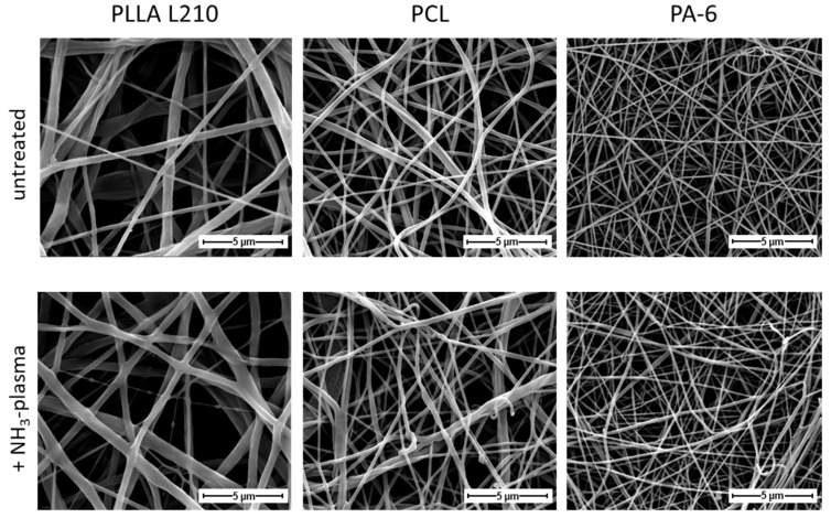
Nanofiber nonwovens are highly promising to serve as biomimetic scaffolds for pioneering cardiac implants such as drug-eluting stent systems or heart valve prosthetics. For successful implant integration, rapid and homogeneous endothelialization is of utmost importance as it forms a hemocompatible surface. This study aims at physicochemical and biological evaluation of various electrospun polymer scaffolds, made of FDA approved medical-grade plastics. Human endothelial cells (EA.hy926) were examined for cell attachment, morphology, viability, as well as actin and PECAM 1 expression. The appraisal of the untreated poly-L-lactide (PLLA L210), poly-ε-caprolactone (PCL) and polyamide-6 (PA-6) nonwovens shows that the hydrophilicity (water contact angle > 80°) and surface free energy (<60 mN/m) is mostly insufficient for rapid cell colonization. Therefore, modification of the surface tension of nonpolar polymer scaffolds by plasma energy was initiated, leading to more than 60% increased wettability and improved colonization. Additionally, NH3-plasma surface functionalization resulted in a more physiological localization of cell−cell contact markers, promoting endothelialization on all polymeric surfaces, while fiber diameter remained unaltered. Our data indicates that hydrophobic nonwovens are often insufficient to mimic the native extracellular matrix but also that they can be easily adapted by targeted post-processing steps such as plasma treatment. The results achieved increase the understanding of cell−implant interactions of nanostructured polymer-based biomaterial surfaces in blood contact while also advocating for plasma technology to increase the surface energy of nonpolar biostable, as well as biodegradable polymer scaffolds. Thus, we highlight the potential of plasma-activated electrospun polymer scaffolds for the development of advanced cardiac implants.

Keywords: CD31; biocompatibility; cardiovascular stent; human endothelial cells; nonwoven.


Conflict of interest statement

The authors declare no conflict of interest.

Figures

Figure 1
Figure 1
Chemical structure of the well-established polymer types for biomedical application used in this study.
Figure 2
Figure 2
Systematic illustration of the study design and methodological background. (I). Preparation of the different used nonwovens using Elmarco nanospider setup, including (1) high-voltage source at the rotating emitter; (2) a bath filled with a polymer solution; (3) fiber formation under solvent evaporation; and (4) a nonwoven collecting unit, (5) which is electrical driven. (II). Surface plasma activation of a nonwoven illustrated by a SEM image. (III). Characterization of the prepared polymer-based nonwovens via comprehensive biological studies, SEM and contact angle measurements, before and after plasma treatment.
Figure 3
Figure 3
High-resolution scanning electron micrographs of morphology of untreated (first row) and NH3-plasma treated (second row) nanofibrous PLLA L210, PCL and PA-6 nonwovens (magnification ×8000, bar = 5 µm).
Figure 4
Figure 4
Fiber diameter of untreated and NH3-plasma functionalized PLLA L210, PCL and PA-6 nonwovens based on individual measurement points. For each polymer, 10 fibers were measured manually in 5 different SEM images (n = 50).
Figure 5
Figure 5
Water contact angle of (A) time-resolved measurement over 60 s using untreated nonwoven material and (B) measurement after a few seconds using untreated and NH3-plasma functionalized PLLA L210, PCL and PA-6 nonwovens.
Figure 6
Figure 6
Relative viability of human endothelial EA.hy926 cells on unmodified and NH3-plasma functionalized polymeric nonwovens after 48 h (mean + SD, n = 6, one-way ANOVA, ns = not significant, and *** p < 0.001).
Figure 7
Figure 7
Cell morphology of human endothelial cells (EA.hy926) on untreated and NH3-plasma functionalized PLLA L210, PCL and PA-6 nonwovens and on a planar control surface (NC) after 48 h (SEM, bar = 40 µm).
Figure 8
Figure 8
Spreading of human endothelial EA.hy926 cells on untreated and NH3-plasma functionalized polymeric nonwovens after 48 h (mean + SD, n = 40, one-way ANOVA, ns = not significant, and *** p < 0.001).
Figure 9
Figure 9
Cell shape described by cell circularity of human endothelial EA.hy926 cells on untreated and NH3-plasma functionalized polymeric nonwovens after 48 h (mean + SD, n = 40, one-way ANOVA, *** p < 0.001).
Figure 10
Figure 10
Fluorescent staining of F-actin in human endothelial EA.hy926 cells grown for 48 h on untreated and NH3-plasma functionalized polymeric nonwovens (red: phalloidin-TRITC for F-actin, blue: Hoechst-staining indicating cell nuclei, confocal microscopy, and bar = 50 μm).
Figure 11
Figure 11
Immunostainings of CD31 in human endothelial EA.hy926 cells on unmodified and NH3-plasma functionalized polymeric nonwovens after 48 h (green: CD31, blue: Hoechst-staining indicating cell nuclei, confocal microscopy, and bar = 50 μm).

References

    1. Mensah G.A., Roth G.A., Fuster V. The Global Burden of Cardiovascular Diseases and Risk Factors: 2020 and Beyond. J. Am. Coll. Cardiol. 2019;74:2529–2532. doi: 10.1016/j.jacc.2019.10.009. - DOI - PubMed
    1. Fioretta E.S., Motta S.E., Lintas V., Loerakker S., Parker K.K., Baaijens F.P.T., Falk V., Hoerstrup S.P., Emmert M.Y. Next-generation tissue-engineered heart valves with repair, remodelling and regeneration capacity. Nat. Rev. Cardiol. 2021;18:92–116. doi: 10.1038/s41569-020-0422-8. - DOI - PubMed
    1. Roshandel M., Dorkoosh F. Cardiac tissue engineering, biomaterial scaffolds, and their fabrication techniques. Polym. Adv. Technol. 2021;32:2290–2305. doi: 10.1002/pat.5273. - DOI
    1. Finn A.V., Joner M., Nakazawa G., Kolodgie F., Newell J., John M.C., Gold H.K., Virmani R. Pathological correlates of late drug-eluting stent thrombosis: Strut coverage as a marker of endothelialization. Circulation. 2007;115:2435–2441. doi: 10.1161/CIRCULATIONAHA.107.693739. - DOI - PubMed
    1. Wessely R. New drug-eluting stent concepts. Nat. Rev. Cardiol. 2010;7:194–203. doi: 10.1038/nrcardio.2010.14. - DOI - PubMed

LinkOut - more resources