

The Cytocompatibility of Silver Diamine Fluoride on Mesenchymal Stromal Cells from Human Exfoliated Deciduous Teeth: An In Vitro Study
- PMID: 35329556
- PMCID: PMC8954535
- DOI: 10.3390/ma15062104
The Cytocompatibility of Silver Diamine Fluoride on Mesenchymal Stromal Cells from Human Exfoliated Deciduous Teeth: An In Vitro Study
Abstract
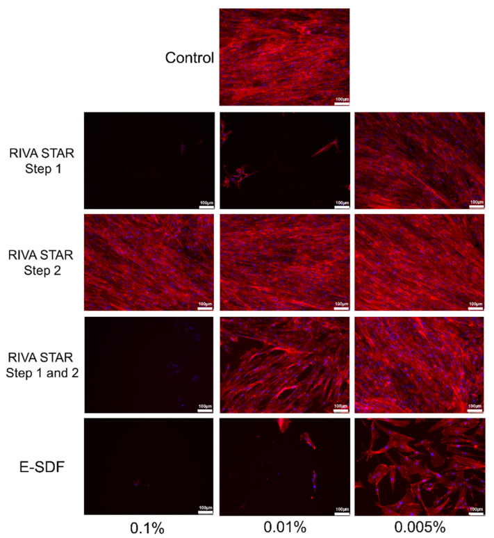
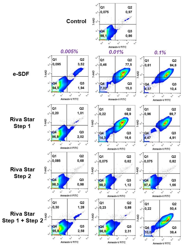
Silver diamine fluoride (SDF) has been used for many years for the treatment of caries, and minimally invasive dentistry concepts have made it popular again. The fact that its application does not require the administration of anesthesia makes its use in children more desirable. The aim of this study was to determine the cytotoxicity of two new commercial SDF products: Riva Star (SDI Dental Limited) and e-SDF (Kids-e-Dental) on mesenchymal stromal cells from human exfoliated deciduous teeth (SHEDs). SHEDs were exposed to SDF products at different concentrations (0.1%, 0.01% and 0.005%). Then different assays were performed to evaluate their cytocompatibility on SHEDs: IC50, MTT, cell migration (wound healing), cell cytoskeleton staining, cell apoptosis, generation of intracellular reactive oxygen species (ROS), and ion chromatography. Statistical analyses were performed using one-way ANOVA and Tukey’s post hoc test (p < 0.05). Riva Star Step 2 showed the same cell metabolic activity when compared to the control condition at any time and concentration. Meanwhile, e-SDF displayed high cytotoxicity at any time and any concentration (*** p < 0.001), whereas Riva Star Step 1 displayed high cytotoxicity at any time at 0.1% and 0.01% (*** p < 0.001). Only e-SDF showed a statistically significant decreased cell migration rate (*** p < 0.001) at all times and in all concentrations. At 0.1%, e-SDF and Riva Star Step 1 only showed 4.37% and 4.47% of viable cells, respectively. These results suggest that Riva Star has better in vitro cytocompatibility on SHEDs than does e-SDF. Riva Star Step 1 was found to be as cytotoxic as e-SDF, but it had better biological properties when mixed with Riva Star Step 2. Our findings suggest that Riva Star is more suitable when used in deciduous teeth due to its lower cytotoxicity compared to e-SDF.
Keywords: SHEDs; cytocompatibility; cytotoxicity; silver diamine fluoride.
Conflict of interest statement
The authors declare that there are no conflict of interest related to this article.
Figures


Similar articles
-
Comparing cytocompatibility of two fluoride-containing solutions and two resin-based restorative materials-a pilot study.Front Oral Health. 2024 Apr 8;5:1330944. doi: 10.3389/froh.2024.1330944. eCollection 2024. Front Oral Health. 2024. PMID: 38650760 Free PMC article.
-
In vitro biocompatibility of ammonia-free silver fluoride products on human dental pulp stem cells.Tissue Cell. 2024 Feb;86:102283. doi: 10.1016/j.tice.2023.102283. Epub 2023 Dec 5. Tissue Cell. 2024. PMID: 38113650
-
Comparison of staining potential of silver diamine fluoride versus silver diamine fluoride and potassium iodide under tooth-colored restorations: An in vitro study.J Indian Soc Pedod Prev Dent. 2021 Jan-Mar;39(1):47-52. doi: 10.4103/jisppd.jisppd_533_20. J Indian Soc Pedod Prev Dent. 2021. PMID: 33885387
-
The use of silver diamine fluoride in a children's hospital: Critical analysis and action protocol.Clin Exp Dent Res. 2022 Oct;8(5):1175-1184. doi: 10.1002/cre2.611. Epub 2022 Jul 22. Clin Exp Dent Res. 2022. PMID: 35869630 Free PMC article. Review.
-
Effect of silver diamine fluoride solution application on the bond strength of dentine to adhesives and to glass ionomer cements: a systematic review.BMC Oral Health. 2020 Feb 5;20(1):40. doi: 10.1186/s12903-020-1030-z. BMC Oral Health. 2020. PMID: 32024501 Free PMC article.
Cited by
-
In Vitro Inhibitory Effect of Silver Diamine Fluoride Combined with Potassium Iodide against Mixed-Species Biofilm Formation on Human Root Dentin.Antibiotics (Basel). 2024 Aug 7;13(8):743. doi: 10.3390/antibiotics13080743. Antibiotics (Basel). 2024. PMID: 39200043 Free PMC article.
-
Effect of Silver Diamine Fluoride Application Methods on Dentin Microhardness and Durability Under pH Cycling: An In Vitro Study.Clin Exp Dent Res. 2025 Aug;11(4):e70190. doi: 10.1002/cre2.70190. Clin Exp Dent Res. 2025. PMID: 40741820 Free PMC article.
-
Comparing cytocompatibility of two fluoride-containing solutions and two resin-based restorative materials-a pilot study.Front Oral Health. 2024 Apr 8;5:1330944. doi: 10.3389/froh.2024.1330944. eCollection 2024. Front Oral Health. 2024. PMID: 38650760 Free PMC article.
-
Pulp cell response to the application of silver diamine fluoride and potassium iodide on caries-like demineralized dentin.Clin Oral Investig. 2023 Dec;27(12):7295-7306. doi: 10.1007/s00784-023-05320-8. Epub 2023 Oct 18. Clin Oral Investig. 2023. PMID: 37853265
References
-
- Paglia L. COVID-19 and Paediatric Dentistry after the lockdown. Eur. J. Paediatr. Dent. 2020;21:89. - PubMed
Grants and funding
LinkOut - more resources
Full Text Sources
