

Retrospective Long-Term Clinical Outcome of Feldspathic Ceramic Veneers
- PMID: 35329602
- PMCID: PMC8954582
- DOI: 10.3390/ma15062150
Retrospective Long-Term Clinical Outcome of Feldspathic Ceramic Veneers
Abstract
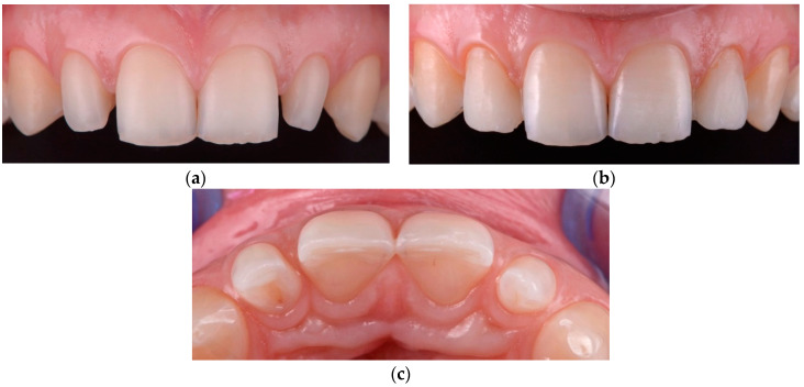
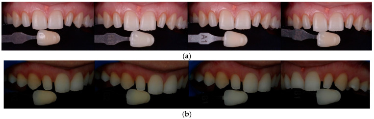
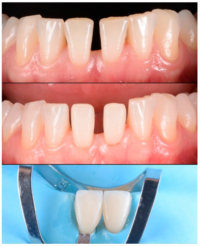
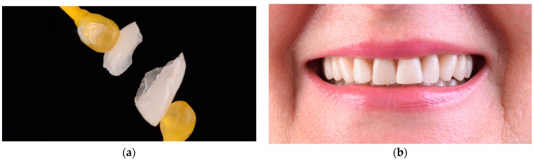
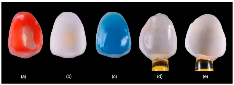
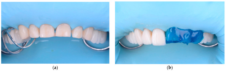
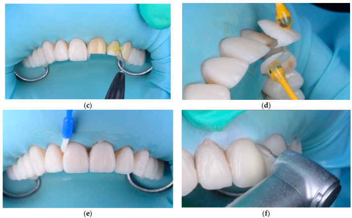
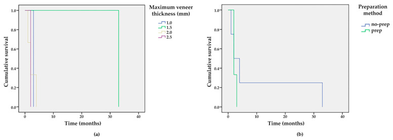
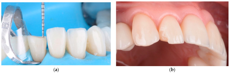
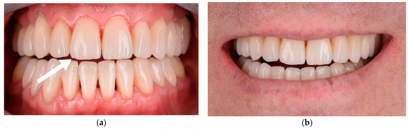
The purpose of this study was to evaluate the clinical outcome of feldspathic ceramic laminate veneers over a 7-year period using minimally invasive techniques, such as vertical preparation (without prosthetic finish line), or no preparation (no-prep). A total of 170 feldspathic ceramic veneers were cemented in the anterior region, including 70 maxillary and 100 mandibular veneers, after special conditioning of the teeth and restorations. The veneers were evaluated using the FDI World Dental Federation criteria evaluation kit after recalling all the patients between February and June 2021. In total, 14 feldspathic veneers failed and were replaced with lithium disilicate because of core fracture, and 10 cases of chipping occurred on the ceramic surface and were polished. The overall survival rate was 91.77% for up to 7 years of function, with a failure rate of 8.23%. In this retrospective survival analysis, the failures, including the fracture of veneers and dental hard tissue, occurred both in prep and no-prep teeth. No failures were observed in veneers with a maximum thickness of 0.5 mm compared to those with a maximum thickness of 1 mm, 1.5 mm, 2 mm, and 2.5 mm.
Keywords: feldspathic ceramic; minimally invasive treatment; no-prep veneers; vertical prep.
Conflict of interest statement
The authors did not have any commercial interest in any of the materials used in this study.
Figures
Similar articles
-
Conventional Versus Minimally Invasive Veneers: A Systematic Review.Cureus. 2023 Sep 4;15(9):e44638. doi: 10.7759/cureus.44638. eCollection 2023 Sep. Cureus. 2023. PMID: 37799216 Free PMC article. Review.
-
Minimally invasive vertical preparation design for ceramic veneers: a multicenter retrospective follow-up clinical study of 265 lithium disilicate veneers.Int J Esthet Dent. 2019;14(3):286-298. Int J Esthet Dent. 2019. PMID: 31312814
-
Combination of minimal- and non-preparation techniques with ceramic veneers for managing esthetic deficiencies.Int J Esthet Dent. 2023 Jul 18;18(3):232-243. Int J Esthet Dent. 2023. PMID: 37462377
-
Performance of ceramic laminate veneers with immediate dentine sealing: An 11 year prospective clinical trial.Dent Mater. 2019 Jul;35(7):1042-1052. doi: 10.1016/j.dental.2019.04.008. Epub 2019 May 10. Dent Mater. 2019. PMID: 31084936
-
A Narrative Review and Clinical Study on Er:YAG Laser Debonding of Ceramic and Composite Veneers.Biomimetics (Basel). 2025 May 6;10(5):295. doi: 10.3390/biomimetics10050295. Biomimetics (Basel). 2025. PMID: 40422124 Free PMC article. Review.
Cited by
-
Challenges faced when masking a single discoloured tooth - Part 2: indirect restoration procedures.Br Dent J. 2025 Jul;239(1):25-30. doi: 10.1038/s41415-025-8385-0. Epub 2025 Jul 11. Br Dent J. 2025. PMID: 40646206 Free PMC article. Review.
-
Conventional Versus Minimally Invasive Veneers: A Systematic Review.Cureus. 2023 Sep 4;15(9):e44638. doi: 10.7759/cureus.44638. eCollection 2023 Sep. Cureus. 2023. PMID: 37799216 Free PMC article. Review.
-
Restoration of Anterior Teeth Defect With Porcelain Laminate Veneer Using the Refractory Technique: A Case Report.Cureus. 2024 Aug 22;16(8):e67501. doi: 10.7759/cureus.67501. eCollection 2024 Aug. Cureus. 2024. PMID: 39310610 Free PMC article.
-
Additive Wax-Up and Diagnostic Mockup As Driving Tools for Minimally Invasive Veneer Preparations.Cureus. 2022 Jul 28;14(7):e27402. doi: 10.7759/cureus.27402. eCollection 2022 Jul. Cureus. 2022. PMID: 36046283 Free PMC article.
-
Retrospective Long-Term Survival Rate and Clinical Performance of Zirconium Oxide Restorations over the Past 5 Years: A Comparative Study Between Single Crowns and Fixed Dental Prostheses.Medicina (Kaunas). 2025 Jan 24;61(2):210. doi: 10.3390/medicina61020210. Medicina (Kaunas). 2025. PMID: 40005327 Free PMC article.
References
-
- Bosch G., Ender A., Mehl A. Non- and minimally invasive full-mouth rehabilitation of patients with loss of vertical dimension of occlusion using CAD/CAM: An innovative concept demonstrated with a case report. Int. J. Comput. Dent. 2015;18:273–286. - PubMed
LinkOut - more resources
Full Text Sources
Miscellaneous
