

Attachment and Osteogenic Potential of Dental Pulp Stem Cells on Non-Thermal Plasma and UV Light Treated Titanium, Zirconia and Modified PEEK Surfaces
- PMID: 35329678
- PMCID: PMC8950369
- DOI: 10.3390/ma15062225
Attachment and Osteogenic Potential of Dental Pulp Stem Cells on Non-Thermal Plasma and UV Light Treated Titanium, Zirconia and Modified PEEK Surfaces
Abstract
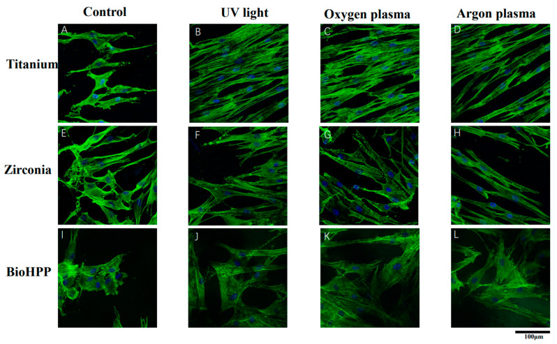
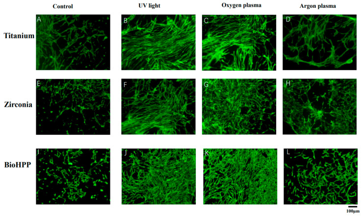
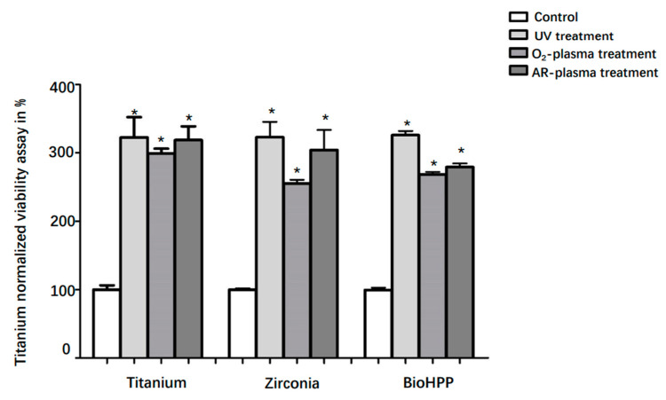
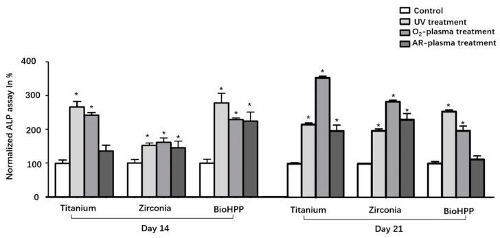
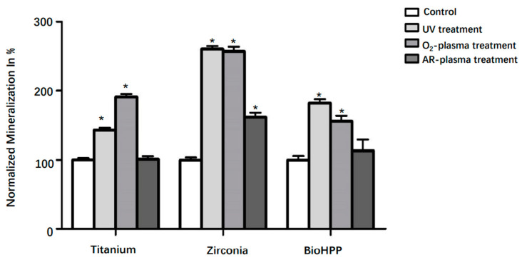
Ultraviolet (UV) light and non-thermal plasma (NTP) treatment are chairside methods that can efficiently improve the biological aging of implant material surfaces caused by customary storage. However, the behaviors of stem cells on these treated surfaces of the implant are still unclear. This study aimed to investigate the effects of UV light and NTP treated surfaces of titanium, zirconia and modified polyetheretherketone (PEEK, BioHPP) on the attachment and osteogenic potential of human dental pulp stem cells (DPSCs) in vitro. Machined disks were treated using UV light and argon or oxygen NTP for 12 min each. Untreated disks were set as controls. DPSCs were cultured from the wisdom teeth of adults that gave informed consent. After 24 h of incubation, the attachment and viability of cells on surfaces were assessed. Cells were further osteogenically induced, alkaline phosphatase (ALP) activity was detected via a p-Nitrophenyl phosphate assay (day 14 and 21) and mineralization degree was measured using a Calcium Assay kit (day 21). UV light and NTP treated titanium, zirconia and BioHPP surfaces improved the early attachment and viability of DPSCs. ALP activity and mineralization degree of osteoinductive DPSCs were significantly increased on UV light and NTP treated surfaces of titanium, zirconia and also oxygen plasma treated Bio-HPP (p < 0.05). In conclusion, UV light and NTP treatments may improve the attachment of DPSCs on titanium, zirconia and BioHPP surfaces. Osteogenic differentiation of DPSCs can be enhanced on UV light and NTP treated surfaces of titanium and zirconia, as well as on oxygen plasma treated Bio-HPP.
Keywords: dental pulp stem cells; modified polyetheretherketone; non-thermal plasma; osteogenesis; titanium; ultraviolet light; zirconia.
Conflict of interest statement
The authors declare no conflict of interest.
Figures
Similar articles
-
Cytocompatibility of Titanium, Zirconia and Modified PEEK after Surface Treatment Using UV Light or Non-Thermal Plasma.Int J Mol Sci. 2019 Nov 8;20(22):5596. doi: 10.3390/ijms20225596. Int J Mol Sci. 2019. PMID: 31717459 Free PMC article.
-
Time Dependency of Non-Thermal Oxygen Plasma and Ultraviolet Irradiation on Cellular Attachment and mRNA Expression of Growth Factors in Osteoblasts on Titanium and Zirconia Surfaces.Int J Mol Sci. 2020 Nov 14;21(22):8598. doi: 10.3390/ijms21228598. Int J Mol Sci. 2020. PMID: 33202662 Free PMC article.
-
Effect of wet storage on the bioactivity of ultraviolet light- and non-thermal atmospheric pressure plasma-treated titanium and zirconia implant surfaces.Mater Sci Eng C Mater Biol Appl. 2019 Dec;105:110049. doi: 10.1016/j.msec.2019.110049. Epub 2019 Aug 2. Mater Sci Eng C Mater Biol Appl. 2019. PMID: 31546363
-
Photofunctionalization and non-thermal plasma activation of titanium surfaces.Clin Oral Investig. 2018 Mar;22(2):1045-1054. doi: 10.1007/s00784-017-2186-z. Epub 2017 Jul 20. Clin Oral Investig. 2018. PMID: 28730456
-
Physicochemical properties of anodized-hydrothermally treated titanium with a nanotopographic surface structure promote osteogenic differentiation in dental pulp stem cells.J Prosthodont Res. 2021 Oct 15;65(4):474-481. doi: 10.2186/jpr.JPR_D_20_00114. Epub 2021 Feb 22. J Prosthodont Res. 2021. PMID: 33612663
Cited by
-
State-of-the-art polyetheretherketone three-dimensional printing and multifunctional modification for dental implants.Front Bioeng Biotechnol. 2023 Oct 19;11:1271629. doi: 10.3389/fbioe.2023.1271629. eCollection 2023. Front Bioeng Biotechnol. 2023. PMID: 37929192 Free PMC article. Review.
-
Innovative Curved-Tip Reactor for Non-Thermal Plasma and Plasma-Treated Water Generation: Synergistic Impact Comparison with Sodium Hypochlorite in Dental Root Canal Disinfection.Materials (Basel). 2023 Nov 17;16(22):7204. doi: 10.3390/ma16227204. Materials (Basel). 2023. PMID: 38005133 Free PMC article.
-
Surface Modifications of High-Performance Polymer Polyetheretherketone (PEEK) to Improve Its Biological Performance in Dentistry.Polymers (Basel). 2022 Dec 16;14(24):5526. doi: 10.3390/polym14245526. Polymers (Basel). 2022. PMID: 36559893 Free PMC article. Review.
-
Antibacterial and Proliferative Effects of NaOH-Coated Titanium, Zirconia, and Ceramic-Reinforced PEEK Dental Composites on Bone Marrow Mesenchymal Stem Cells.Pharmaceutics. 2022 Dec 28;15(1):98. doi: 10.3390/pharmaceutics15010098. Pharmaceutics. 2022. PMID: 36678727 Free PMC article.
-
Effect of different agents on preload force of dental implants with bio high-performance poly-ether-ether-ketone abutments.J Oral Biol Craniofac Res. 2024 Nov-Dec;14(6):756-760. doi: 10.1016/j.jobcr.2024.10.004. Epub 2024 Oct 17. J Oral Biol Craniofac Res. 2024. PMID: 39493258 Free PMC article.
References
-
- Sullivan R.M. Implant dentistry and the concept of osseointegration: A historical perspective. J. Calif. Dent. Assoc. 2001;29:737–745. - PubMed
-
- Merli M., Migani M., Bernardelli F., Esposito M. Vertical bone augmentation with dental implant placement: Efficacy and complications associated with 2 different techniques. A retrospective cohort study. Int. J. Oral Maxillofac. Implant. 2006;21:600–606. - PubMed
Grants and funding
LinkOut - more resources
Full Text Sources
