. 2022 Mar 18;15(6):2246.
doi: 10.3390/ma15062246.

A Preliminary In Vitro Study of 3D Full-Field Strain Distribution in Human Whole Premolars Using Digital Image Correlation

Affiliations

A Preliminary In Vitro Study of 3D Full-Field Strain Distribution in Human Whole Premolars Using Digital Image Correlation

Qing Liu et al. Materials (Basel). .

Abstract

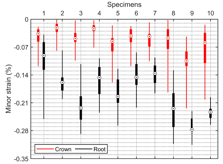
Full-field measurements can provide a more complete description of the behavior of human whole tooth under load. To that end, in vitro experiments were carried out to measure the full-field buccal surface strains of human premolars free of caries and abrasion using digital image correlation (DIC). Experimental results show that both the value field and the orientation field of strains can be observed exactly, both of which contain a wealth of information. Furthermore, the strain distributions between the crown and the root of specimens were significantly different (p < 0.001). An interesting observation was a watershed at the cementoenamel junction (CEJ) which separates the orientation field of strains into two distinct parts; the watershed was also observed in the value field of strains in some specimens whose geometries changed obviously at the CEJ. Another interesting observation was that the minor strains increased linearly from cervical to apical regions in the root cementum. Experimental results also support the viewpoint that mechanisms of non-carious cervical lesions (NCCLs) may in part be due to the changing orientation of tensile strains, as well as their magnitude, and they also support the hypothesis that occlusal force can contribute to root fractures.

Keywords: buccal surface strain; digital image correlation; full-field measurement; human premolars.


Conflict of interest statement

The authors declare no conflict of interest.

Figures

Figure 1
Figure 1
Some prepared specimens in cardboard boxes.
Figure 2
Figure 2
Measurement setup. The 3D DIC device monitoring the buccal surface of the specimen that fixed on the fixture of the testing machine.
Figure 3
Figure 3
Calculation of full-field buccal surface strains in the software of 3D DIC device. The image marked “0” at the bottom left corner is the image captured by the left camera of the 3D DIC device, while the image marked “1” is the image captured by the right camera of the 3D DIC device. Virtual grids (green square grids) were down-sampled for display purposes.
Figure 4
Figure 4
Full-field buccal surface strains of the specimen (shown in Figure 3) at (a) stage 0, (b) stage 5, (c) stage 10, (d) stage 20, (e) stage 30, and (f) stage 39. The first row displays the major strain (or tensile strain) map, while the second row displays the minor strain (or compressive strain) map.
Figure 5
Figure 5
Stage–strain curves of three representative points (POINT1, POINT2, and POINT3, as shown in Figure 3) at the center line on the buccal surface of the specimen. The trend lines were fitted according to the experimental data.
Figure 6
Figure 6
The orientation fields of the minor strains in Figure 4. (a) Specimen. (bf) Stage 5, stage 10, stage 20, stage 30, and stage 39, respectively. The short thin black lines (unit vector), mapped onto the value fields, represent the directions of minor strains, while the purple dotted lines represent the location of the CEJ.
Figure 7
Figure 7
The orientation distributions of the minor strains of (a) another specimen at (b) stage 15 and (c) stage 30. The purple dotted lines denote the CEJ. It can be seen that the watershed exists in both the orientation field and the value field of minor strains.
Figure 8
Figure 8
Section line along the center line of the value field of minor strains at stage 39 in Figure 4.
Figure 9
Figure 9
Box plot of the major strains in the crown and the root of specimens. Boxes are composed of the minimum, first quartile, third quartile, median, and maximum strain values.
Figure 10
Figure 10
Box plot of the minor strains in the crown and the root of specimens. The composition of the boxes is the same as described in Figure 9.

Similar articles

References

    1. Thompson V.P. The tooth: An analogue for biomimetic materials design and processing. Dent. Mater. 2020;36:25–42. doi: 10.1016/j.dental.2019.08.106. - DOI - PubMed
    1. Shahar R., Weiner S. Insights into whole bone and tooth function using optical metrology. J. Mater. Sci. 2007;42:8919–8933. doi: 10.1007/s10853-007-1693-8. - DOI
    1. Arola D.D., Gao S., Zhang H., Masri R. The Tooth: Its Structure and Properties. Dent. Clin. N. Am. 2017;61:651–668. doi: 10.1016/j.cden.2017.05.001. - DOI - PMC - PubMed
    1. Zhang Y.R., Du W., Zhou X.D., Yu H.Y. Review of research on the mechanical properties of the human tooth. Int. J. Oral Sci. 2014;6:61–69. doi: 10.1038/ijos.2014.21. - DOI - PMC - PubMed
    1. Picart P., Fages M., Slangen P., Xia H., Montresor S., Guo R., Li J., Solieman O.Y., Durand J.C. A review on optical methods to assess dental behavior under stress; Proceedings of the SPIE, Optical Methods for Inspection, Characterization, and Imaging of Biomaterials IV; Munich, Germany. 21 June 2019.

LinkOut - more resources