. 2022 Mar 19;15(6):2274.
doi: 10.3390/ma15062274.

Devising Bone Molecular Models at the Nanoscale: From Usual Mineralized Collagen Fibrils to the First Bone Fibers Including Hydroxyapatite in the Extra-Fibrillar Volume

Affiliations

Devising Bone Molecular Models at the Nanoscale: From Usual Mineralized Collagen Fibrils to the First Bone Fibers Including Hydroxyapatite in the Extra-Fibrillar Volume

Amadeus C S Alcântara et al. Materials (Basel). .

Abstract

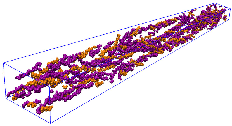
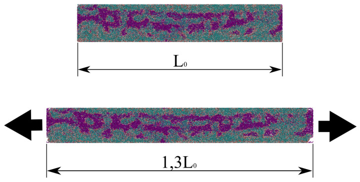
At the molecular scale, bone is mainly constituted of type-I collagen, hydroxyapatite, and water. Different fractions of these constituents compose different composite materials that exhibit different mechanical properties at the nanoscale, where the bone is characterized as a fiber, i.e., a bundle of mineralized collagen fibrils surrounded by water and hydroxyapatite in the extra-fibrillar volume. The literature presents only models that resemble mineralized collagen fibrils, including hydroxyapatite in the intra-fibrillar volume only, and lacks a detailed prescription on how to devise such models. Here, we present all-atom bone molecular models at the nanoscale, which, differently from previous bone models, include hydroxyapatite both in the intra-fibrillar volume and in the extra-fibrillar volume, resembling fibers in bones. Our main goal is to provide a detailed prescription on how to devise such models with different fractions of the constituents, and for that reason, we have made step-by-step scripts and files for reproducing these models available. To validate the models, we assessed their elastic properties by performing molecular dynamics simulations that resemble tensile tests, and compared the computed values against the literature (both experimental and computational results). Our results corroborate previous findings, as Young's Modulus values increase with higher fractions of hydroxyapatite, revealing all-atom bone models that include hydroxyapatite in both the intra-fibrillar volume and in the extra-fibrillar volume as a path towards realistic bone modeling at the nanoscale.

Keywords: bone elastic properties; bone nanoscale model; collagen fiber; extra-fibrillar volume; hydroxyapatite; mineralized collagen fibril; molecular dynamics.


Conflict of interest statement

The authors declare no conflict of interest.

Figures

Figure A1
Figure A1
Model of human triple-helix CLG structure devised by homology modeling using MODELLER and shown in VMD with drawing and coloring methods Quicksurf and Chain, respectively.
Figure A2
Figure A2
Chains A of the lowest DOPE model built with allhmodel (blue) and 3HR2 (red) are shown in VMD as VDW. Zoomed (top) and distant (bottom) views.
Figure A3
Figure A3
Extraction of CLG NanoFiber (blue) from the seven-by-seven periodic replication (orange transparent) of the CLG Fibril (red). View of xz-plane (left) and yz-plane (right) in VMD.
Figure A4
Figure A4
CLG Fiber (blue): a two-by-two replication of CLG NanoFiber, seen among the seven-by-seven periodic replication of the CLG Fibril’s UC (orange transparent). View of xz-plane (left) and yz-plane (right) in VMD.
Figure A5
Figure A5
Devised CLG Fiber shown in VMD. The CLG chains (three chains each in one color) of a previous specific CLG NanoFiber are all represented five times (in the overlap zone) by the same three colors, e.g., five CLG molecules with their three chains in gray, cyan, and black. The different replications of the previous CLG NanoFiber can be identified by the colors cyan, red, yellow, and orange.
Figure 1
Figure 1
Structural representation of the backbone of a single molecule (top) and fibril (bottom) of the type I CLG. Chains A and C (alpha-1) are indicated in the purple color, and Chain B (alpha-2) in orange one.
Figure 2
Figure 2
Schematic view of the modeling of a structure that resembles fibers in bones.
Figure 3
Figure 3
CLG Fibril within UC; snapshot from VMD viewer.
Figure 4
Figure 4
CLG Fibril periodically replicated in space; snapshot from VMD viewer.
Figure 5
Figure 5
Bone Fiber view of the xy-plane (VMD). The simulation box (blue) defines the external boundary of the EFV. The IFV box (red) defines the external boundary of the IFV and the internal boundary of the EFV. Only CLG backbone molecules are shown.
Figure 6
Figure 6
Bone Fiber view (VMD) of yz-plane (top) and xz-plane (bottom). The simulation box (blue) defines the external boundary of the EFV. The IFV box (red) defines the external boundary of the IFV and the internal boundary of the EFV. Only CLG backbone molecules are shown.
Figure 7
Figure 7
Simulation box of a Bone Fiber model, and a view of a 3-by-3 periodic replication of its xy-cross-section (VMD). HA, H2O and CLG molecules are shown in cyan, red, and purple (alpha-1 chains) and orange colors (alpha-2 chains), respectively.
Figure 8
Figure 8
Zoomed view of a bone fiber in VMD. HA, H2O and CLG molecules are shown in cyan, red, and purple (alpha-1 chains) and orange colors (alpha-2 chains), respectively.
Figure 9
Figure 9
RMSD of Bone Fiber models (with respect to devised models, frame 0).
Figure 10
Figure 10
Bone Fiber 55 UC before (top) and after (bottom) the tensile test; snapshots from OVITO [66]. The arrows indicate the stretching directions.
Figure 11
Figure 11
Stress–strain curves computed for the devised models, and their respective linear regression.

Similar articles

Cited by

References

    1. Odén A., McCloskey E., Kanis J., Harvey N.C., Johansson H. Burden of high fracture probability worldwide: Secular increases 2010–2040. Osteoporos. Int. 2015;26:2243–2248. doi: 10.1007/s00198-015-3154-6. - DOI - PubMed
    1. Alcantara A.C.S., Assis I., Prada D., Mehle K., Schwan S., Costa-Paiva L., Skaf M.S., Wrobel L.C., Sollero P. Patient-Specific Bone Multiscale Modelling, Fracture Simulation and Risk Analysis—A Survey. Materials. 2020;13:106. doi: 10.3390/ma13010106. - DOI - PMC - PubMed
    1. Sabet F.A., Raeisi Najafi A., Hamed E., Jasiuk I. Modelling of bone fracture and strength at different length scales: A review. Interface Focus. 2016;6:20150055. doi: 10.1098/rsfs.2015.0055. - DOI - PMC - PubMed
    1. Sherman V.R., Yang W., Meyers M.A. The materials science of collagen. J. Mech. Behav. Biomed. Mater. 2015;52:22–50. doi: 10.1016/j.jmbbm.2015.05.023. - DOI - PubMed
    1. Mohammad M.-G., Wang X.Z. Nanomechanics and Ultrastructure of Bone: A Review. Comput. Model. Eng. Sci. 2020;125:1–32. doi: 10.32604/cmes.2020.012123. - DOI

LinkOut - more resources