Risk Factors, Clinical and Endoscopic Features, and Clinical Outcomes in Patients with Cytomegalovirus Esophagitis
- PMID: 35329909
- PMCID: PMC8955160
- DOI: 10.3390/jcm11061583
Risk Factors, Clinical and Endoscopic Features, and Clinical Outcomes in Patients with Cytomegalovirus Esophagitis
Abstract
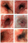
Cytomegalovirus (CMV) esophagitis is the second most common CMV disease of the gastrointestinal tract. This study aims to comprehensively analyze risk factors, clinical characteristics, endoscopic features, outcomes, and prognostic factors of CMV esophagitis. We retrospectively collected data of patients who underwent esophageal CMV immunohistochemistry (IHC) staining between January 2003 and April 2021 from the pathology database at the Chang Gung Memorial Hospital. Patients were divided into the CMV and non-CMV groups according to the IHC staining results. We enrolled 148 patients (44 CMV and 104 non-CMV patients). The risk factors for CMV esophagitis were male sex, immunocompromised status, and critical illness. The major clinical presentations of CMV esophagitis included epigastric pain (40.9%), fever (36.4%), odynophagia (31.8%), dysphagia (29.5%), and gastrointestinal bleeding (29.5%). Multiple diffuse variable esophageal ulcers were the most common endoscopic feature. The CMV group had a significantly higher in-hospital mortality rate (18.2% vs. 0%; p < 0.001), higher overall mortality rate (52.3% vs. 14.4%; p < 0.001), and longer admission duration (median, 24 days (interquartile range (IQR), 11−47 days) vs. 14 days (IQR, 7−24 days); p = 0.015) than the non-CMV group. Acute kidney injury (odds ratio (OR), 174.15; 95% confidence interval (CI), 1.27−23,836.21; p = 0.040) and intensive care unit admission (OR, 26.53; 95% CI 1.06−665.08; p = 0.046) were predictors of in-hospital mortality. In conclusion, the mortality rate of patients with CMV esophagitis was high. Physicians should be aware of the clinical and endoscopic characteristics of CMV esophagitis in high-risk patients for early diagnosis and treatment.
Keywords: acute kidney injury; cytomegalovirus; endoscopy; esophagitis; prognostic factor.
Conflict of interest statement
The authors declare no conflict of interest relevant to this manuscript.
Figures


Similar articles
-
Clinical manifestations, risk factors, and prognostic factors of cytomegalovirus enteritis.Gut Pathog. 2021 Aug 18;13(1):53. doi: 10.1186/s13099-021-00450-4. Gut Pathog. 2021. PMID: 34407879 Free PMC article.
-
Cytomegalovirus gastritis: Clinicopathological profile.Dig Liver Dis. 2021 Jun;53(6):722-728. doi: 10.1016/j.dld.2020.12.002. Epub 2021 Jan 11. Dig Liver Dis. 2021. PMID: 33441265
-
The clinical characteristics and manifestations of cytomegalovirus esophagitis.Dis Esophagus. 2016 May;29(4):392-9. doi: 10.1111/dote.12340. Epub 2015 Feb 26. Dis Esophagus. 2016. PMID: 25715747
-
Surgical manifestations of gastrointestinal cytomegalovirus infection in children: Clinical audit and literature review.J Pediatr Surg. 2015 Nov;50(11):1874-9. doi: 10.1016/j.jpedsurg.2015.06.018. Epub 2015 Jun 30. J Pediatr Surg. 2015. PMID: 26265193 Review.
-
Optimal Management of Patients with Phlegmonous Esophagitis: A Systematic Review and Meta-Analysis.J Clin Med. 2023 Nov 17;12(22):7147. doi: 10.3390/jcm12227147. J Clin Med. 2023. PMID: 38002759 Free PMC article. Review.
Cited by
-
IgG4-Related Oesophageal Disease with Cytomegalovirus Infection: A Case Report.J Pers Med. 2023 Mar 9;13(3):493. doi: 10.3390/jpm13030493. J Pers Med. 2023. PMID: 36983676 Free PMC article.
-
Clinical Characteristics of Cytomegalovirus Disease of the Upper Gastrointestinal Tract: A 10-Year Multicenter Retrospective Study.Korean J Helicobacter Up Gastrointest Res. 2023 Dec;23(4):294-301. doi: 10.7704/kjhugr.2023.0054. Epub 2023 Dec 8. Korean J Helicobacter Up Gastrointest Res. 2023. PMID: 40503509 Free PMC article.
-
Infective Esophagitis.Korean J Helicobacter Up Gastrointest Res. 2025 Jun;25(2):108-116. doi: 10.7704/kjhugr.2025.0007. Epub 2025 Jun 4. Korean J Helicobacter Up Gastrointest Res. 2025. PMID: 40550542 Free PMC article. Review. English.
-
Viral esophagitis in non-human immunodeficiency virus patients: a case-control study.Transl Gastroenterol Hepatol. 2024 Mar 29;9:19. doi: 10.21037/tgh-23-44. eCollection 2024. Transl Gastroenterol Hepatol. 2024. PMID: 38716211 Free PMC article.
-
Esophagitis in a Post-Liver Transplant Patient: A Case of Cytomegalovirus and Herpes Simplex Virus-1 Coinfection.Clin Case Rep. 2024 Nov 19;12(11):e9565. doi: 10.1002/ccr3.9565. eCollection 2024 Nov. Clin Case Rep. 2024. PMID: 39568531 Free PMC article.
References
-
- Li L., Chakinala R.C. StatPearls. StatPearls Publishing LLC.; Treasure Island, FL, USA: 2021. Cytomegalovirus Esophagitis. - PubMed
LinkOut - more resources
Full Text Sources
Miscellaneous

