. 2022 Mar 16;11(6):1660.
doi: 10.3390/jcm11061660.

Hypersensitivity Myocarditis after COVID-19 mRNA Vaccination

Affiliations

Hypersensitivity Myocarditis after COVID-19 mRNA Vaccination

Andrea Frustaci et al. J Clin Med. .

Abstract

Background: Myocarditis, even in a severe and lethal form, may occur after COVID-19 mRNA (BNT162b2) vaccination. However, its pathway, morphomolecular characterization and treatment are still unknown.

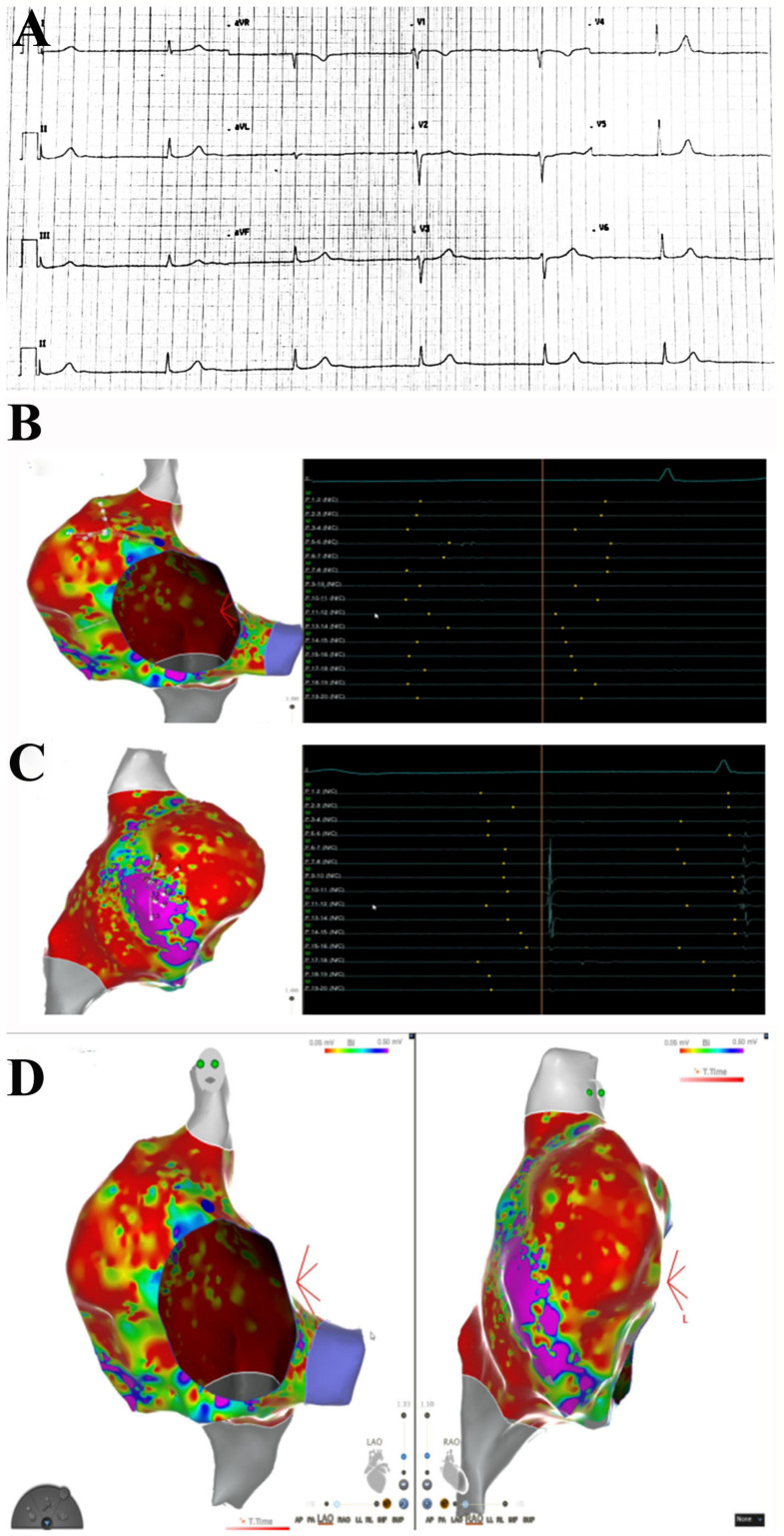
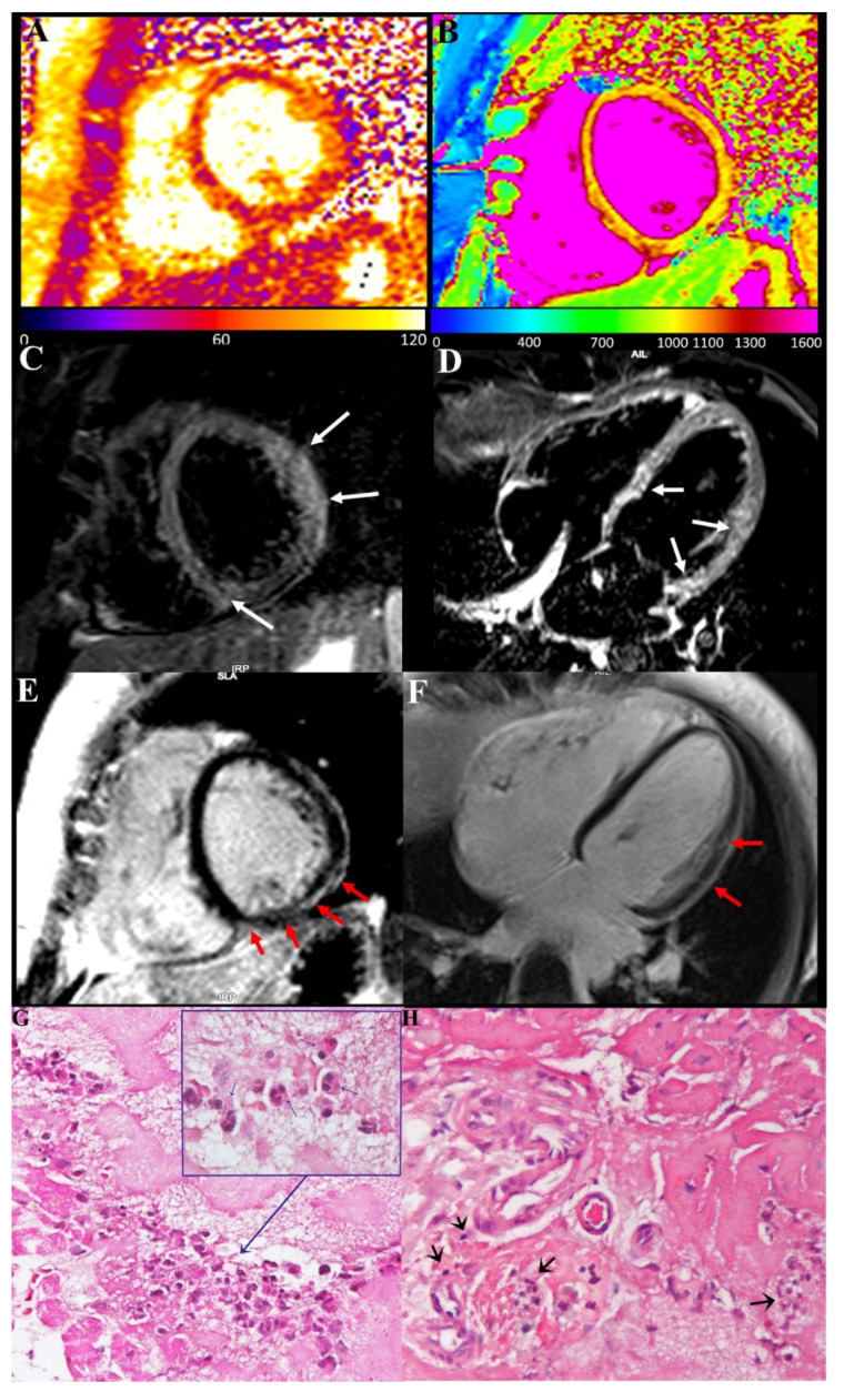
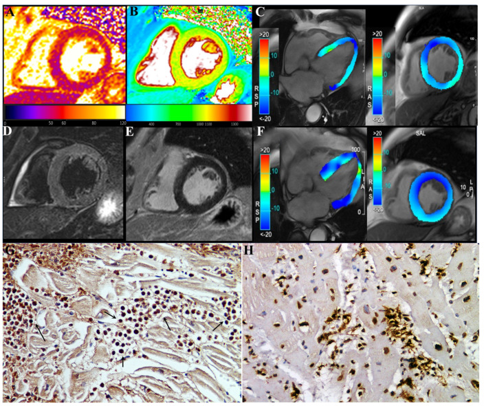
Methods: Routine hematochemical screening, ECG, Holter monitoring, 2D echocardiogram cardiac magnetic resonance (CMR) and invasive cardiac studies (cardiac catheterization, selective coronary angiography, left ventriculography and left ventricular endomyocardial biopsy) are reported from three patients (39F-pt1, 78M-pt2, 52M-pt3) with severe compromise of conduction tissue (junctional rhythm and syncope, pt1) or cardiac function compromise (LVEF ≤ 35%, pt2 and pt3) after COVID-19 mRNA (BNT162b2).

Results: Hematochemical data and coronary angiography were normal in the patients studied. Histology showed in all three patients extensive myocardial infiltration of degranulated eosinophils and elevation of serum cationic protein directly responsible for cardiomyocyte damage. These findings demonstrate myocarditis hypersensitivity to some component of the vaccine (spike protein?) acting as a hapten to some macromolecules of cardiomyocytes. Steroid administration (prednisone, 1 mg/kg die for 3 days, followed by 0.33 mg/kg for 4 weeks) was followed by complete recovery of cardiac contractility in pt2 and pt3.

Conclusions: Eosinophilic myocarditis is a possible adverse reaction to the mRNA COVID-19 vaccine. Its pathway is mediated by release of cationic protein and responds to short courses of steroid administration.

Keywords: COVID-19 mRNA vaccination; eosinophilic myocarditis; myocarditis.


Conflict of interest statement

The authors declare that they have no competing interest.

Figures

Figure 1
Figure 1
Right atrial substrate mapping and endocavitary electrogram recordings. ECG shows no sinus rhythm, no atrial activity at EPS. Junctional rhythm at 40 bts/min (A). Endocavitary recordings show the absence of atrial activation during atrial mapping (B). Some atrial signals are recorded on the anterolateral wall of right atrium (C). Left and right anterior oblique projections of right atrium endocardial substrate mapping shows a large scarred area with low-voltage signals (<0.05 mV, in red), with a small portion of myocardial wall with preserved voltage signals (>0.5 mV, in purple) on the anterolateral aspect of right atrium (D).
Figure 2
Figure 2
Eosinophilic myocarditis with conduction tissue involvement after COVID-19 mRNA vaccination. (A,B): Myocardial T2 (A) and T1 (B) maps, acquired on mid-ventricular short-axis view, showed a diffuse increase in both T2 and T1 myocardial value as a result of a diffuse inflammatory involvement. (C,D): Presence of myocardial edema was also confirmed on STIR T2-weighted images acquired on short-axis (C) and horizontal long-axis (D) views as multiple areas of hyperintense signal within the basal septum, the inferior interventricular junction and mid-basal anterolateral wall (arrows). (E,F): Late-enhancement images acquired on the same short-axis (E) and horizontal long-axis (F) planes reveal tiny areas of mild enhancement with “patchy” distribution and predominant involvement of subepicardial layer in the same location of the above-mentioned myocardial edema (red arrows). (G,H): Histology shows degranulation of crystalloids of eosinophilic granulocytes (see insert and blue arrow) responsible for the release of the cationic protein and ultimately for myocardial damage and a strongly infiltration of eosinophils in the conduction tissue (H) (black arrow). (400× magnification).
Figure 3
Figure 3
A 78-year-old male (pt2) with eosinophilic myocarditis myocarditis after second dose of COVID-19 mRNA (BNT162b2) vaccine. At first CMR exam (AE), performed 4 weeks after symptoms onset, T2 (A) and native T1 (B) maps revealed an increase in global T2 (55 ms ± 4 ms, normal range < 49.9 ms) and T1 (1060 ± 15 ms, normal range < 1027 ms) myocardial values, without any noticeable focal areas of signal changes on STIR T2-weighted (D) and late-gadolinium-enhanced (E) images, reflecting mild diffuse edematous condition. CineMR images, acquired on four-chamber (left) and mid-ventricular short-axis (right) views, analyzed by tissue tracking analysis show moderate impairment of both longitudinal and circumferential systolic left ventricular function (GLS: −12.3%, GCS: −13.6%, EF: 35%) at admission (C), which improved at two-month follow-up after therapy ((F), GLS: −18.1%, GCS: −17.8%, EF: 49.5%). GCS: global circumferential strain; GLS: global longitudinal strain; EF: ejection fraction. (G) shows in post-vax myocarditis massive eosinophilic myocardial infiltration with necrosis of the adjacent myocytes (IHC for eosinophil major basic protein (EMBP) antibody). (200× magnification). (H): Comparison with acute myocarditis associated to COVID19 infection showing intense lymphocytic inflammation (IHC for CD45Ro) (200× magnification).

Similar articles

Cited by

References

    1. Mevorach D., Anis E., Cedar N., Bromberg M., Haas E.J., Nadir E., Olsha-Castell S., Arad D., Hasin T., Levi N., et al. Myocarditis after BNT162b2 mRNA Vaccine against Covid-19 in Israel. N. Engl. J. Med. 2021;385:2140–2149. doi: 10.1056/NEJMoa2109730. - DOI - PMC - PubMed
    1. Husby A., Hansen J.V., Fosbøl E., Thiesson E.M., Madsen M., Thomsen R.W., Sørensen H.T., Andersen M., Wohlfahrt J., Gislason G., et al. SARS-CoV-2 vaccination and myocarditis or myopericarditis: Population based cohort study. BMJ. 2021;375:e068665. doi: 10.1136/bmj-2021-068665. - DOI - PMC - PubMed
    1. Verma A.K., Lavine K.J., Lin C.-Y. Myocarditis after Covid-19 mRNA Vaccination. N. Engl. J. Med. 2021;385:1332–1334. doi: 10.1056/NEJMc2109975. - DOI - PMC - PubMed
    1. Aretz H.T., Billingham M.E., Edwards W.D., Factor S.M., Fallon J., Fenoglio J.J., Olsen E.G., Schoen F.J. Myocarditis. A histopathologic definition and classification. Am. J. Cardiovasc. Pathol. 1987;1:3–14. - PubMed
    1. Caforio A.L., Pankuweit S., Arbustini E., Basso C., Gimeno-Blanes J., Felix S.B., Fu M., Heliö T., Heymans S., Jahns R., et al. Current state of knowledge on aetiology, diagnosis, management, and therapy of myocarditis: A position statement of the European Society of Cardiology Working Group on Myocardial and Pericardial Diseases. Eur. Heart J. 2013;34:2636–2648. doi: 10.1093/eurheartj/eht210. - DOI - PubMed

LinkOut - more resources